
Fistulogram and Fistuloplasty
- Home
- Fistulogram and Fistuloplasty
What is an Arteriovenous (AV) Fistula?
An arteriovenous (AV) fistula is a surgically created connection between an artery and a vein, usually in the arm, for hemodialysis access. It is considered the best vascular access because it provides:
- High blood flow for dialysis
- Lower infection risk
- Longer lifespan compared to catheters and grafts
Over time, stenosis (narrowing), thrombosis (clotting), or other complications may develop, reducing blood flow. To diagnose and treat these problems, fistulogram and fistuloplasty are performed.
FISTULOGRAM
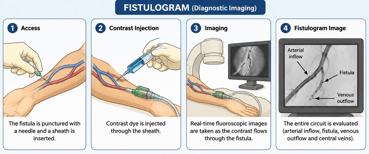
- A fistulogram (also called fistula angiography) is a diagnostic imaging procedure used to evaluate the structure and blood flow of an AV fistula or AV graft.
- A contrast dye is injected into the fistula while X-ray images are taken to identify abnormalities.
Indications for Fistulogram
- A fistulogram is performed when there are signs of fistula dysfunction, such as:
Clinical Signs
- Difficult cannulation
- Increased venous pressure during dialysis
- Decreased dialysis adequacy
- Reduced blood flow rate
- Prolonged bleeding after needle removal
- Weak or absent thrill
- Abnormal bruit
- Arm swelling
- Suspected stenosis
- Suspected thrombosis
- Recirculation during dialysis
Pre-Procedure Assessment
1.Clinical Evaluation
- Review dialysis records
- Examine thrill and bruit
- Assess arm swelling
- Check previous interventions
2. Laboratory Tests
- CBC
- PT/INR
- Platelet count
- Kidney function tests (if needed)
Patient Preparation
- Explain procedure
- Obtain informed consent
- Fasting for 2 to 4 hours
- Review medications
- Assess allergy history
- Establish IV access if required
Findings on Fistulogram
-Normal Findings
- Smooth vessel walls
- Good blood flow
- No narrowing
- Patent central veins
-Abnormal Findings
- Stenosis
- Narrowing of vessel lumen.
- Thrombosis-Partial clot/ Complete clot
- Aneurysm- Localized vessel dilatation
- Accessory Veins- Side branches diverting blood flow
- Central Venous Stenosis-Narrowing of subclavian or brachiocephalic veins
FISTULOPLASTY
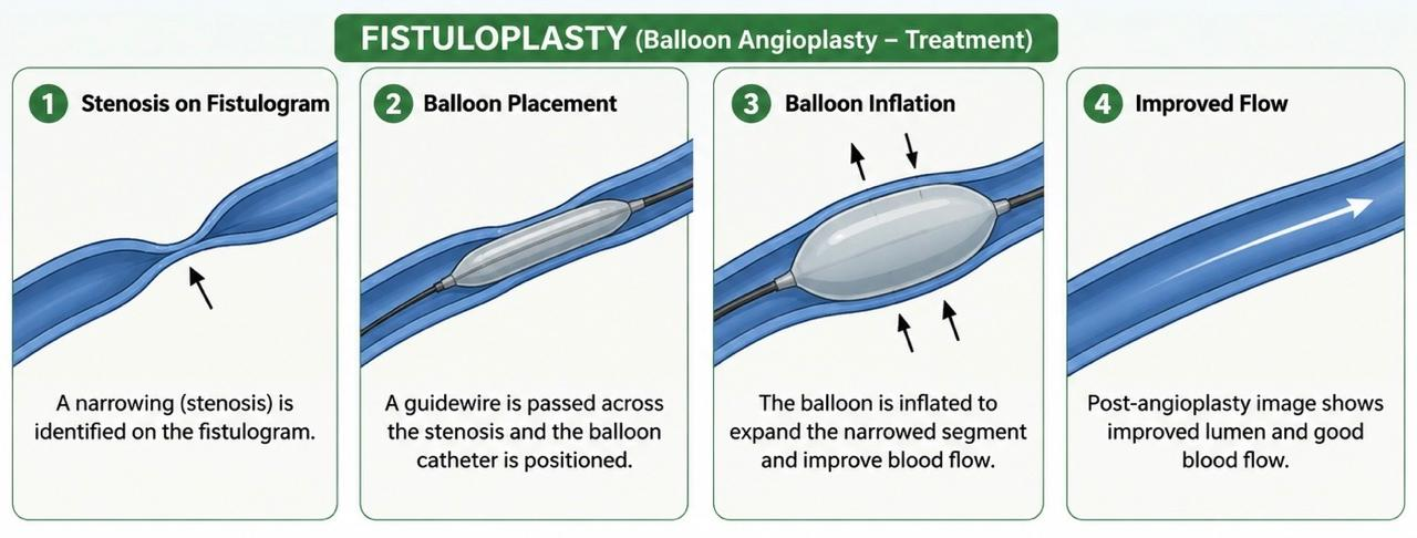
- Fistuloplasty (Percutaneous Transluminal Angioplasty – PTA) is a minimally invasive procedure used to treat stenosis detected during fistulogram.
- A balloon catheter is inflated inside the narrowed segment to widen the vessel and restore blood flow.
Indications
Significant Stenosis
-Usually:
- 50% narrowing
- Associated with clinical dysfunction
Other Indications
- Poor dialysis flow
- Elevated venous pressures
- Arm swelling
- Access recirculation
- Recurrent thrombosis
Types of Fistuloplasty
1. Standard Balloon Angioplasty
- Uses conventional balloon.
- Most common method.
2. High-Pressure Balloon Angioplasty- Used for resistant lesions.
-Balloon inflation pressure:
- 20–30 atmospheres or higher
3. Cutting Balloon Angioplasty
Balloon contains tiny blades.
Used for:
- Fibrotic lesions
- Recurrent stenosis
4. Drug-Coated Balloon Angioplasty- Balloon coated with antiproliferative medication.
- May reduce restenosis.
Procedure of Fistulogram:
The patient is positioned comfortably on the fluoroscopy table with the access arm exposed. The skin over the AV fistula is thoroughly cleaned using an antiseptic solution to reduce the risk of infection. Sterile drapes are then placed around the procedure site to maintain a sterile field throughout the examination.
After preparing the site, local anesthesia is administered by infiltrating lidocaine into the skin and surrounding tissues at the planned puncture site. This helps minimize pain and discomfort during the procedure.
Once adequate anesthesia is achieved, the AV fistula is punctured using a needle under sterile conditions. A small vascular sheath is then inserted into the fistula, providing access for contrast injection and catheter manipulation if required.
A radiographic contrast dye is injected through the sheath into the fistula. As the contrast flows through the vascular access, real-time fluoroscopic (X-ray) imaging is performed to visualize the blood vessels and assess blood flow patterns.
A series of fluoroscopic images are obtained to evaluate the entire dialysis access circuit. The examination includes assessment of the arterial inflow, the arteriovenous anastomosis, the body of the fistula, the venous outflow tract, and the central veins. These images help identify any stenosis, thrombosis, aneurysm formation, or other abnormalities that may affect the function of the dialysis access.
Post Procedure Care:
Immediate Monitoring
- Vital signs
- Access site bleeding
- Thrill and bruit assessment
Post Procedure Care Patient Instructions:
- Keep dressing dry
- Avoid heavy lifting for 24 hours
- Report swelling or bleeding
- Observe for infection
Dialysis
- Most patients can continue with dialysis using the fistula within 24 hours or even the same day if advised.
Success Rate
- Technical Success- 85–95%
Clinical Success
- Significant improvement in dialysis performance
- Reduced venous pressures
- Better dialysis adequacy
SUMMARY:
- Fistulogram (The Mapping) = Look at the fistula.
- Fistuloplasty (The Repair) = Open the narrowed fistula.
Would you like to request an appointment?
You can call on +91-98200 86520 for Appointments or fill the form below