
Brain Aneurysm Coiling
- Home
- Brain Aneurysm Coiling
What is brain aneurysm?
-A brain aneurysm is a balloon-like swelling that results from a weakness in the wall of one of the arteries supplying blood to the brain.
-If the aneurysm ruptures (bursts), it causes bleeding around the brain called a subarachnoid haemorrhage (SAH), which is a life-threatening emergency associated with stroke, brain damage, or even death.
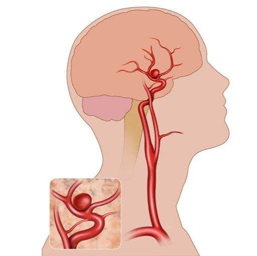
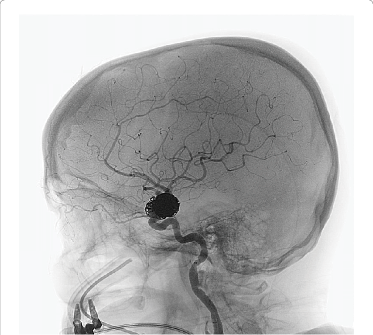
Locations of Brain Aneurysms
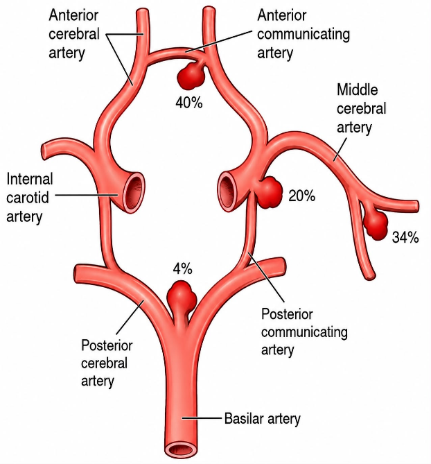
-Brain aneurysms most commonly occur at branching points of the arteries at the base of the brain, particularly around the Circle of Willis.
The most common sites are:
- Anterior communicating artery (ACoA)
- Internal carotid artery (ICA)
- Posterior communicating artery (PCoA)
- Middle cerebral artery (MCA)
- Posterior cerebral artery (PCA)
- Basilar artery tip
- Posterior circulation arteries
-These locations are important when planning cerebral angiography and endovascular coiling, because the aneurysm’s location, size, neck, and relationship to nearby branches determine the appropriate treatment technique
What is brain aneurysm coiling?
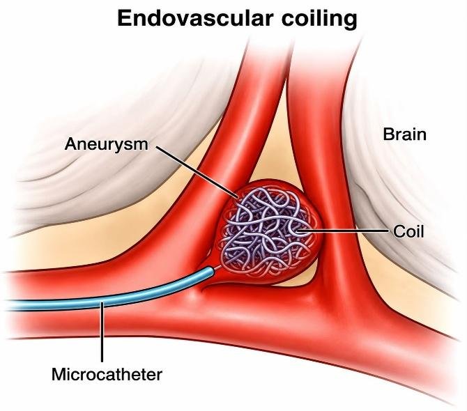
-Brain aneurysm coiling is a minimally invasive endovascular procedure performed by an interventional radiologist or interventional neuroradiologist.
-Instead of opening the skull, the doctor inserts a catheter through an artery in the wrist or groin and places tiny platinum coils inside the aneurysm. Compared to conventional open surgery, brain aneurysm coiling offers faster healing, lower complication rates in selected cases, and excellent long-term outcomes. Today, many patients diagnosed with aneurysms are being treated successfully using this advanced endovascular coiling technique.
These coils:
- Slow blood flow into the aneurysm
- Promote blood clot formation inside the aneurysm
- Seal off the aneurysm from the circulation
- Reduce the risk of rupture or rebleeding
The normal artery remains open while the aneurysm is closed.
Goals of Coiling
- Prevent aneurysm rupture
- Prevent rebleeding after rupture
- Preserve normal blood flow to the brain
- Reduce the risk of stroke
- Avoid open brain surgery when appropriate
Who Needs Brain Aneurysm Coiling?
Coiling may be recommended for:
1. Ruptured aneurysms- These require emergency treatment because bleeding has already occurred.
Symptoms include:
- Sudden severe headache (“worst headache of life”)
- Vomiting
- Neck stiffness
- Loss of consciousness
- Seizures
- Weakness
- Confusion
2. Unruptured aneurysms
Treatment may be recommended if:
- Large aneurysm
- Growing aneurysm
- Irregular shape
- Family history of aneurysms
- Symptoms caused by pressure on nearby nerves
- High risk of rupture
Small aneurysms without high-risk features may simply be monitored.
When is Coiling Not Suitable?
Coiling may not be the best option if:
- The aneurysm has a very wide neck
- The aneurysm shape is unsuitable
- Important arteries arise from the aneurysm
- The aneurysm is extremely large or complex
In these cases, alternatives include:
- Surgical clipping
- Balloon-assisted coiling
- Stent-assisted coiling
- Flow diverter placement
Pre-Procedure Evaluation
1. Medical History
-The doctor asks about-Previous stroke, high blood pressure, diabetes, smoking, headaches, previous aneurysm, heart disease, kidney disease, allergies to contrast dye, blood-thinning medicines, previous surgeries
2. Physical Examination
-The doctor checks:
A thorough physical examination is performed to assess your general health and neurological function before brain aneurysm coiling.
- Blood pressure: Your blood pressure is measured because high blood pressure increases the risk of aneurysm rupture and should be controlled before treatment.
- Heart rate: Your heart rate is checked to ensure your heart is functioning normally before the procedure.
- Neurological examination: The doctor evaluates your brain and nervous system to identify any existing neurological problems and establish a baseline for comparison after the procedure. Which includes muscle strength, speech, vision, balance, cranial nerves.
3. Blood Tests
-These may include:
- Complete Blood Count (CBC): This test measures your red blood cells, white blood cells, and platelets to check for anaemia, infection, or bleeding problems.
- Kidney Function Tests: These tests assess how well your kidneys are working because they help remove the contrast dye used during the procedure.
- Liver Function Tests: These tests evaluate the health of your liver, which plays an important role in metabolism and blood clotting.
- Blood Clotting Tests (PT/INR and aPTT): These tests determine how well your blood clots and help identify any increased risk of bleeding during the procedure.
- Blood Sugar: Your blood glucose level is checked because well-controlled blood sugar reduces the risk of complications and promotes better recovery.
- Blood Group and Cross-Match (if required): This test identifies your blood type and prepares compatible blood in case a blood transfusion is needed, although this is uncommon during brain aneurysm coiling.
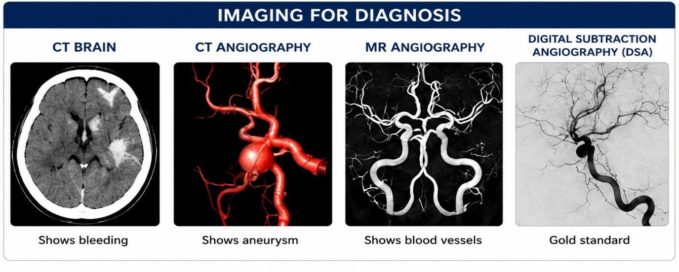
4.Imaging Tests
- Non-contrast CT Brain : Looks for bleeding in the brain.
- CT Angiography (CTA) : Shows the aneurysm and nearby blood vessels.
- MRI Brain : Assesses brain tissue and any previous injury.
- MR Angiography (MRA) : Provides detailed images of brain arteries.
- Digital Subtraction Angiography (DSA) : This is the gold standard for diagnosing brain aneurysms and planning treatment.
Preparation Before the Procedure
- Do not eat or drink for several hours (if advised).
- Inform your doctor about all medications.
- Blood thinners may need adjustment.
- Remove jewellery, dentures, and contact lenses.
- An IV line is inserted.
- Consent is obtained
Step-by-Step Procedure
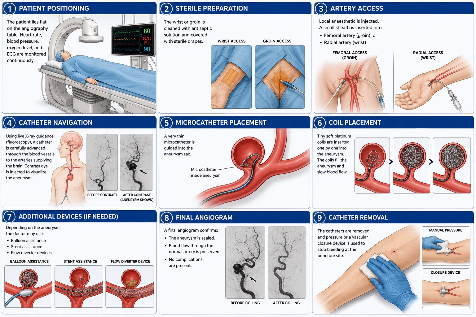
The patient lies flat on the angiography table. Heart rate, blood pressure, oxygen level, and ECG are monitored continuously.
-The wrist or groin is cleaned with antiseptic solution and covered with sterile drapes.
-Local anaesthetic is injected.
-A small sheath is inserted into:
- Femoral artery (groin), or
- Radial artery (wrist)
-Using live X-ray guidance (fluoroscopy), a catheter is carefully advanced through the blood vessels to the arteries supplying the brain.
-Contrast dye is injected to visualize the aneurysm.
-A very thin microcatheter is guided into the aneurysm sac.
-Tiny soft platinum coils are inserted one by one into the aneurysm.
-The coils fill the aneurysm and slow blood flow
-Depending on the aneurysm, the doctor may use:
- Balloon assistance
- Stent assistance
- Flow diverter devices
-A final angiogram confirms:
- The aneurysm is sealed.
- Blood flow through the normal artery is preserved.
- No complications are present.
-The catheters are removed, and pressure or a vascular closure device is used to stop bleeding at the puncture site.
After the Procedure
-The patient is monitored in the recovery area or intensive care unit (especially after a ruptured aneurysm).
-Monitoring includes:
- Blood pressure
- Heart rate
- Oxygen level
- Neurological status
- Puncture site
Recovery
Most people can:
- Walk within 24 hours
- Resume light activities in about 1–2 weeks
- Avoid heavy lifting for several weeks or as advised
Follow-Up
-Follow-up imaging is important because some aneurysms may reopen over time.
-Your doctor may recommend:
- MR Angiography (MRA)
- CT Angiography (CTA)
- Digital Subtraction Angiography (DSA)
Benefits
- Minimally invasive
- No open brain surgery
- Smaller incision
- Faster recovery
- Reduced hospital stay
- Effective treatment for many aneurysms
- Lower risk of wound complications
Risks and Complications
Although generally safe, complications can include:
- Stroke
- Bleeding
- Rupture of the aneurysm during the procedure
- Blood clots
- Coil migration
- Narrowing of the artery
- Contrast allergy
- Kidney injury from contrast (rare)
- Infection
- Groin or wrist bleeding
- Recurrence or reopening of the aneurysm, requiring follow-up
Lifestyle After Coiling
- Take prescribed medications as directed.
- Control high blood pressure.
- Stop smoking.
- Maintain a healthy weight.
- Exercise regularly
- Eat a balanced diet.
- Attend all follow-up appointments.
Would you like to request an appointment?
You can call on +91-98200 86520 for Appointments or fill the form below