
Dural Arteriovenous Fistula Embolization
- Home
- Dural Arteriovenous Fistula Embolization
What is an Arteriovenous Malformation (AVM)?
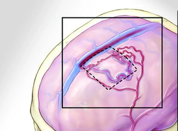
A Dural Arteriovenous Fistula Embolization is a minimally invasive, image-guided procedure used to block the abnormal connection between arteries and veins in the brain or spinal cord’s protective outer covering called The Dura Mater. It is performed by an interventional neuroradiologist or neurointerventional surgeon.
A thin catheter is inserted into an artery and guided to the fistula. A special material is injected to block the abnormal connection and restore normal blood flow.
The procedure avoids open brain surgery in many patients.
The procedure is performed under X-ray guidance (Digital Subtraction Angiography – DSA).
Goals of Embolization
The aim of embolization is to:
- Completely close the abnormal connection
- Reduce abnormal blood flow
- Prevent brain haemorrhage
- Improve neurological symptoms
- Reduce pressure inside abnormal veins
- Preserve normal brain functioning
- Protect the brain from future damage
What is a Dural Arteriovenous Fistula (dAVF)?
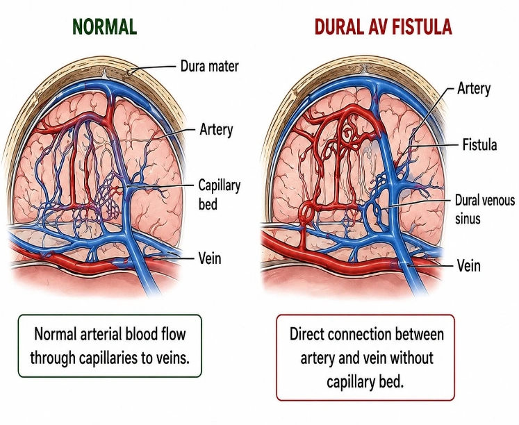
-A Dural Arteriovenous Fistula (dAVF) is an abnormal connection between arteries and veins located within the dura mater, the tough outer covering of the brain or spinal cord.
Normally:
- Arteries carry oxygen-rich blood to the brain.
- Blood passes through tiny capillaries.
- Veins carry blood back to the heart.
In a dAVF:
- The arteries connect directly to the veins without capillaries.
- Blood flows under high pressure into veins.
- This can cause enlarged veins, bleeding, stroke, seizures, or neurological symptoms.
-This abnormal pressure can lead to:
- Brain swelling
- Bleeding (brain haemorrhage)
- Stroke-like symptoms
- Neurological problem
What Does “Dural” Mean?
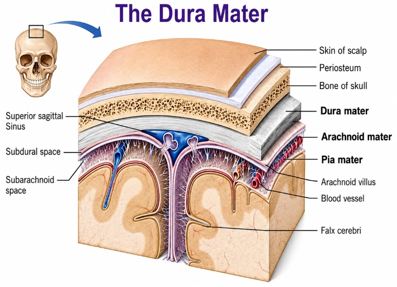
-The brain is covered by three protective layers (meninges):
- Dura mater (outer layer)
- Arachnoid mater
- Pia mater (inner layer)
-A dural AV fistula occurs within the dura mater, unlike a brain AVM (arteriovenous malformation), which occurs inside the brain tissue.
Common Intracranial Sites for dAVM
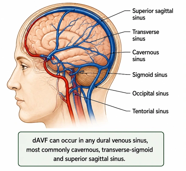
- Transverse-sigmoid sinus: Found at the back and side of the head; the single most frequent site.
- Cavernous sinus: Located near the base of the brain behind the eyes, often causing eye-related symptoms.
- Superior sagittal sinus: Runs along the top center of the head.
- Straight sinus: Located centrally within the tentorial folds inside the brain
- Tentorial region – less common but can have high-risk venous drainage.
Who Needs Embolization?
-Embolization is recommended when:
- The fistula has cortical venous drainage (high-risk dAVF)
- There is brain bleeding
- The patient has neurological symptoms
- Pulsatile tinnitus is severe
- Vision problems occur
- Progressive neurological deficits develop
- Seizures occur
- Imaging shows a high risk of haemorrhage
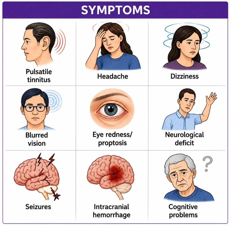
Symptoms of dAVF
Symptoms depend on the location.
Common symptoms include:
- Pulsating sound in the ear (pulsatile tinnitus- a whooshing sound)
- Headache
- Dizziness
- Blurred or double vision
- Eye swelling or redness
- Weakness of an arm or leg
- Difficulty speaking
- Numbness
- Seizures
- Memory problems
- Brain haemorrhage
- Difficulty walking
Causes and Risk Factors
Often the exact cause is unknown.
Possible causes include:
- Previous head injury
- Dural venous sinus thrombosis
- Previous brain surgery
- Brain infection
- Aging
- Rarely congenital abnormalities
Pre-Procedure Evaluation
1. Medical History : A doctor asks about symptoms such as headaches, tinnitus (a whooshing sound in the ear), previous stroke, seizures, any history of bleeding, allergies specially to contrast dye, previous surgeries, and wheather you are taking any blood thinning medicines. This helps assess your overall health and plan the safest treatment
2. Physical Examination : A complete neurological examination is performed to assess vision, speech, balance, muscle strength, sensation, reflexes, and cranial nerve function. This helps identify any neurological problems caused by the dural AV fistula.
3. Blood Tests :
- Complete blood count (CBC)- This test measures your red blood cells, white blood cells, and platelets to check for anaemia, infection, or bleeding problems.
- Kidney function tests– These tests assess how well your kidneys are working because they help remove the contrast dye used during the procedure.
- Liver function tests– These tests evaluate the health of your liver, which plays an important role in metabolism and blood clotting.
- Coagulation Profile (PT/INR and aPTT)- These tests determine how well your blood clots and help identify any increased risk of bleeding during the procedure.
- Blood Sugar– Your blood glucose level is checked because well-controlled blood sugar reduces the risk of complications and promotes better recovery.
- Blood Group and Cross-Match (if required)- This test identifies your blood type and prepares compatible blood in case a blood transfusion is needed, although this is uncommon during brain aneurysm coiling.
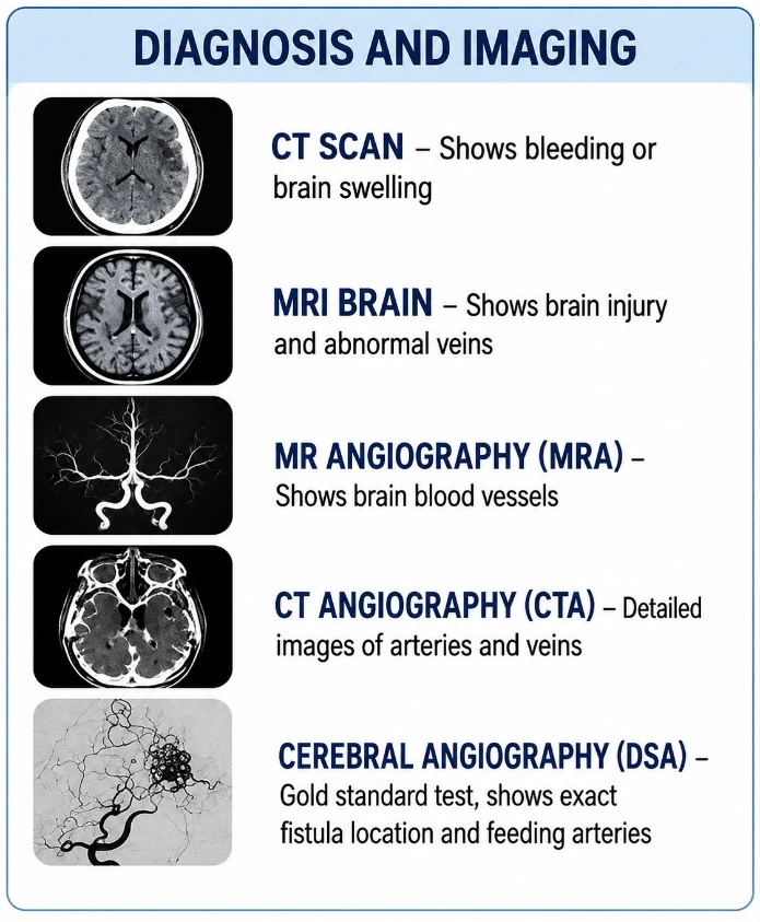
4. Imaging Tests:
- CT Scan-Looks for bleeding or brain swelling.
- MRI Brain– Shows brain injury and abnormal veins.
- MR Angiography (MRA)– Shows blood vessels and blood flow.
- CT Angiography (CTA)- Provides detailed images of arteries and veins.
- Cerebral Angiography (Digital Subtraction Angiography – DSA)
This is the gold standard investigation.
It helps identify:
- Feeding arteries
- Exact fistula location
- Draining veins
- Venous reflux
- Number of arterial feeders
- Best treatment approach
Preparation Before the Procedure
- Fast for about 6–8 hours (if instructed).
- Take regular medicines only as advised.
- Stop blood thinners if instructed.
- Remove jewellery and dentures.
- Wear a hospital gown.
- An intravenous (IV) line is inserted.
- Consent is obtained after discussing benefits and risks.
Step-by-Step Embolization Procedure
-The patient lies supine (flat on back) on the angiography table.
-Heart rate, blood pressure, oxygen level, and ECG are continuously monitored.
-General anaesthesia is commonly used, although selected cases may be done under conscious sedation.
-The groin (femoral artery) or wrist (radial artery) is cleaned with antiseptic solution and covered with sterile drapes.
-Local anaesthetic is injected to numb the skin at the access site.
-A small needle punctures the artery. A vascular sheath is inserted.
-Using continuous X-ray (fluoroscopy), the doctor advances a guiding catheter through the blood vessels to the arteries supplying the fistula.
-Contrast dye is injected.
-Multiple angiographic images identify:
- Feeding arteries
- Exact fistula
- Venous drainage
- Dangerous branches
- Normal arteries that must be preserved
-A very thin microcatheter is advanced through the guiding catheter and positioned as close to the fistula as possible.
-The doctor slowly injects an embolic agent to block the abnormal connection.
Common embolic materials include:
- Onyx (ethylene-vinyl alcohol copolymer) – the most commonly used liquid embolic agent.
- PHIL (Precipitating Hydrophobic Injectable Liquid).
- n-BCA (n-butyl cyanoacrylate) – medical glue.
- Coils (used in selected cases, especially through the venous approach).
-The embolic material fills the fistula and blocks the abnormal blood flow while preserving normal vessels.
-Another angiogram is performed to confirm:
- Complete closure of the fistula (when possible)
- Normal blood flow to the brain
- No significant residual shunting
-The catheters and sheath are removed.
-Pressure or a vascular closure device is used to stop bleeding.
-The patient is moved to the recovery room or intensive care unit for observation.
After the Procedure
Patients are monitored for:
- Blood pressure
- Neurological status
- Headache
- Bleeding from the puncture site
- Limb circulation
- Allergic reactions
A follow-up CT scan or MRI may be performed if needed.
Recovery at Home
Patients should:
- Drink plenty of fluids (unless restricted).
- Avoid heavy lifting for about one week.
- Keep the puncture site clean and dry.
- Take medicines as prescribed.
- Attend follow-up appointments.
Benefits of Embolization
- Minimally invasive
- No large surgical incision
- Shorter hospital stay
- Faster recovery
- High success rate in appropriately selected patients
- Can be curative in many cases
- Lower risk than open surgery for many fistulas
Possible Risks and Complications
Although generally safe, complications can include:
- Bleeding at the puncture site
- Infection
- Stroke
- Brain haemorrhage
- Temporary or permanent neurological deficits
- Injury to normal blood vessels
- Contrast allergy
- Kidney injury from contrast (rare)
- Incomplete closure of the fistula
- Recurrence requiring further treatment
Follow-Up
Patients usually require:
- Neurological examinations
- MRI or MRA
- Follow-up cerebral angiography (DSA), often performed months after treatment to confirm complete closure
- Additional embolization if residual fistula remains
Other Treatment Options for Dural AV Fistula (Short)
1. Microsurgical Disconnection
- Open brain surgery is performed to disconnect the abnormal draining vein.
- Usually recommended when embolization is not feasible or has failed.
- Often provides a permanent cure.
2. Stereotactic Radiosurgery (Gamma Knife/CyberKnife)
- Focused radiation is delivered to the fistula.
- The abnormal vessels gradually close over 1–3 years.
- Best suited for selected small or residual dAVFs that are not at immediate high risk of bleeding.
3. Combined Treatment
- A combination of embolization with surgery or radiosurgery may be used for complex fistulas to improve the chance of complete cure.
4. Observation
- Some low-risk dAVFs without dangerous venous drainage and with minimal symptoms may be monitored with regular clinical and imaging follow-up instead of immediate treatment.
Frequently Asked Questions (FAQs)
1. Is embolization a major surgery?
- No. It is a minimally invasive procedure performed through a small puncture in the artery.
2. Is the procedure painful?
- No. It is usually performed under general anaesthesia, so you do not feel pain during the procedure.
3. Can the fistula come back after treatment?
- Most patients are cured, but some may have residual or recurrent fistulas, which is why follow-up imaging is important.
Would you like to request an appointment?
You can call on +91-98200 86520 for Appointments or fill the form below