
Carotid Artery Stenting
- Home
- Carotid Artery Stenting
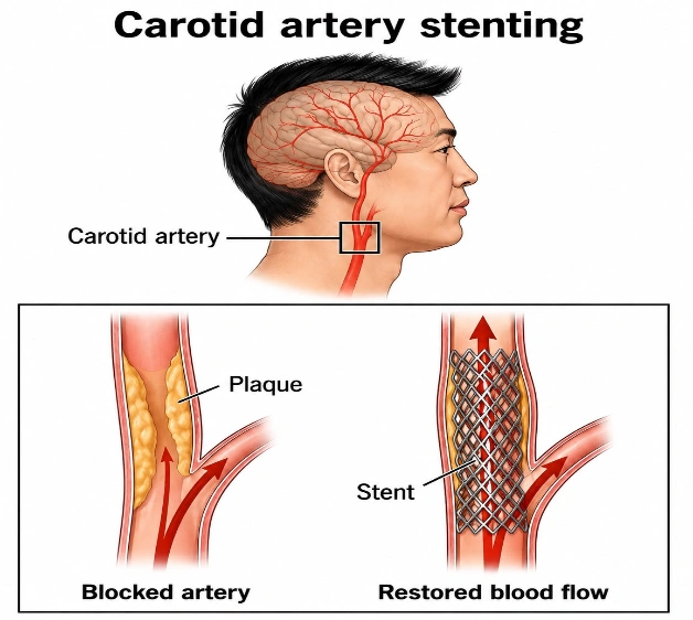
Carotid Artery Angioplasty and Stenting (CAS) is a minimally invasive procedure used to treat narrowing (stenosis) of the carotid arteries The carotid arteries are the two main arteries located on either side of the neck that supply oxygen-rich blood to the brain.
When fatty deposits (plaque) build up inside these arteries, blood flow to the brain decreases, increasing the risk of a transient ischemic attack (TIA) or stroke
-During CAS, an interventional radiologist or vascular specialist:
- Inserts a thin catheter through an artery (usually in the groin or wrist).
- Inflates a tiny balloon to widen the narrowed artery (angioplasty).
- Places a metal mesh stent to keep the artery open.
- Uses a cerebral protection device to catch debris and reduce the risk of stroke during the procedure.
Anatomy of the Carotid Arteries
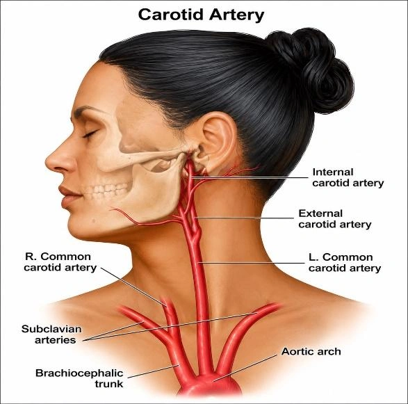
-There are two carotid arteries:
- Right Common Carotid Artery- Arises from the brachiocephalic artery.
- Left Common Carotid Artery- Arises directly from the aortic arch.
-Each common carotid artery divides into:
- Internal carotid artery (ICA): Supplies the brain.
- External carotid artery (ECA): Supplies the face, scalp, and neck.
-Most carotid narrowing occurs at the carotid bifurcation and the beginning of the internal carotid artery.
What is Carotid Artery Stenosis?
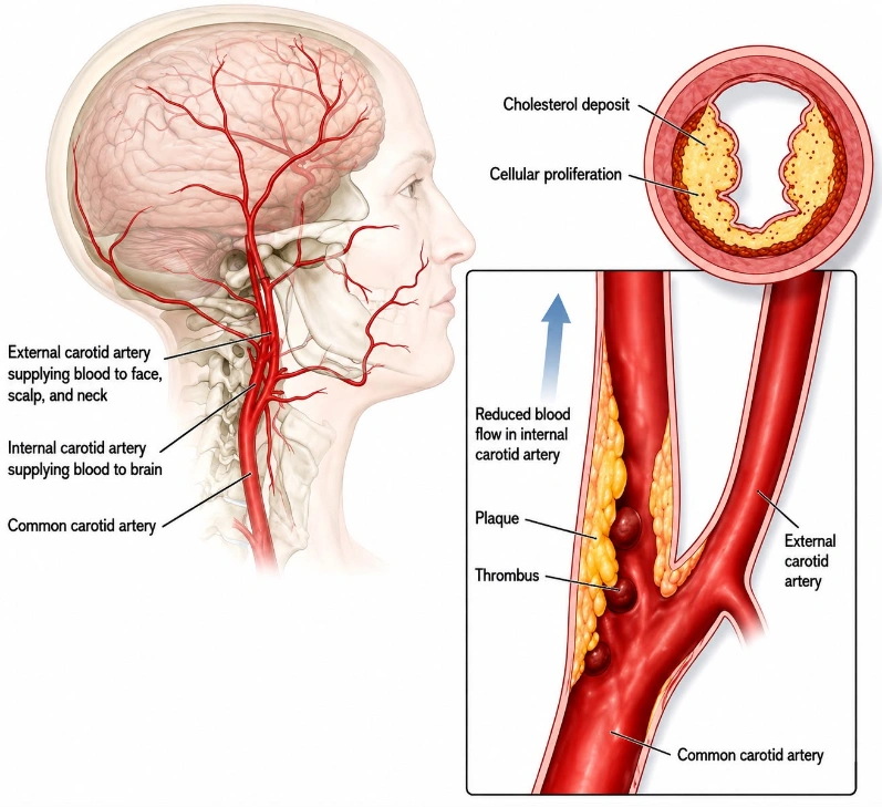
Carotid artery stenosis means narrowing of the carotid artery due to plaque buildup.
-Plaque contains:
- Cholesterol
- Fat
- Calcium
- Inflammatory cells
- Fibrous tissue
Over time, plaque narrows the artery and can break off, sending clots to the brain and causing a stroke
Causes:
The most common cause is atherosclerosis.
Other causes include:
- High blood pressure
- High cholesterol
- Diabetes
- Smoking
- Increasing age
- Obesity
- Lack of exercise
- Family history
- Radiation to the neck
- Fibromuscular dysplasia (rare)
Risk Factors:
- Age over 60 years
- Smoking
- Diabetes
- High cholesterol
- High blood pressure
- Coronary artery disease
- Peripheral arterial disease
- Previous stroke or TIA
- Chronic kidney disease
- Obesity
Symptoms:
Many people have no symptoms until the artery becomes severely narrowed.
-Symptoms may include:
- Sudden weakness of the face, arm, or leg (usually on one side)
- Difficulty speaking
- Slurred speech
- Vision loss in one eye
- Dizziness
- Difficulty walking
- Loss of balance
- Sudden confusion
–These symptoms require immediate medical attention.
Indications for Carotid Artery Stenting
-CAS may be recommended in patients with:
- Symptomatic carotid artery stenosis
- Significant asymptomatic carotid stenosis in selected patients
- High surgical risk for carotid endarterectomy
- Previous neck surgery or radiation
- Restenosis after carotid endarterectomy
- High carotid artery lesions that are difficult to reach surgically
Contraindications
-CAS may not be suitable if the patient has:
- Complete carotid artery occlusion (blockage)
- Active bleeding disorder
- Untreated infection
- Severe allergy to contrast dye that cannot be managed
- Severe kidney failure (requires careful planning)
- Inability to take antiplatelet medications
- Very tortuous or heavily calcified arteries making catheter passage unsafe
Pre-Procedure Evaluation
1. CLINICAL ASSESSMENT
-The doctor evaluates:
- Symptoms– such as weakness, numbness, difficulty speaking, or temporary loss of vision.
- History of TIA (transient ischemic attack) or stroke– this may indicate significant carotid artery disease.
- Heart disease– also reviewed before the procedure.
- Kidney functions– particularly because contrast dye may be used during angiography.
- Medical history– such as previous surgeries, current medications, and known allergies, especially allergies to contrast material or medications.
2. PHYSICAL EXAMINATION
- Neurological examination– a detailed neurological examination is performed to check speech, vision, muscle strength, sensation, coordination, and other neurological functions
- Blood pressure is also measured.
- Heart examination– to assess the heart rate and rhythm and to look for any signs of cardiovascular disease.
- Neck examination– the neck is examined, including listening for a carotid bruit, which may suggest turbulent blood flow through a narrowed carotid artery.
- Pulse assessment– the doctor also checks the patient’s pulses to assess circulation.
3. BLOOD TESTS
- Complete blood count (CBC)- to check the levels of red blood cells, white blood cells, and platelets.
- Kidney function tests– serum creatinine and eGFR, are done to determine whether the kidneys can safely handle contrast dye.
- Coagulation profile (PT/INR, aPTT)- checks how well the blood clots and helps assess bleeding risk.
- Blood sugar– particularly to identify or monitor diabetes
4. IMAGING TESTS
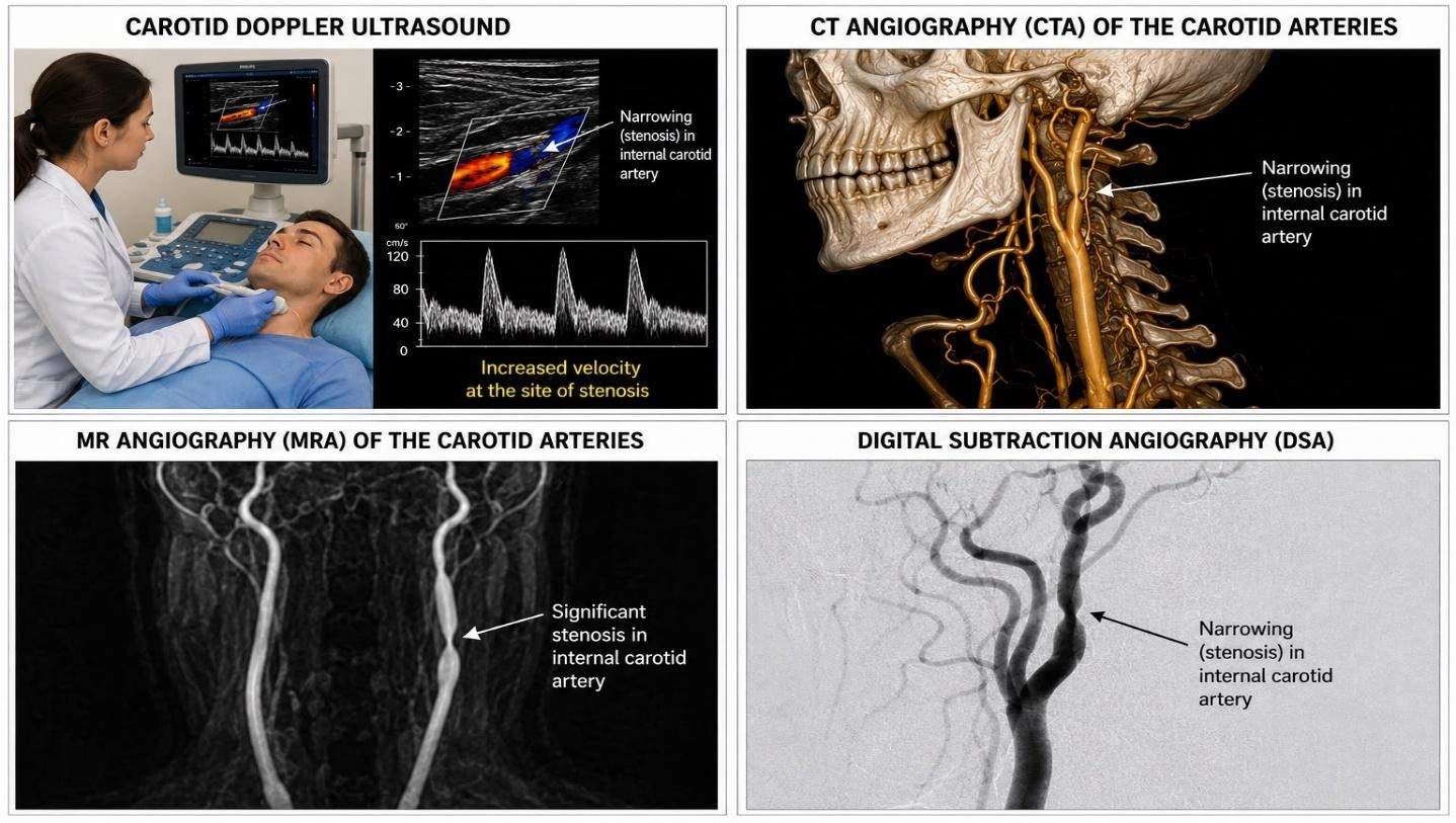
- Carotid Doppler Ultrasound- First-line test to assess blood flow and estimate degree of narrowing.
- CT Angiography (CTA)– Shows the severity and location of stenosis.
- MR Angiography (MRA)– Provides detailed images without ionizing radiation.
- Diagnostic Cerebral Angiography– Gold standard. Performed when more detailed vascular mapping is required. Often performed immediately before stenting.
Patient Preparation
- Fast for 2 hours as advised by the doctor.
- Continue or stop medications only as instructed.
- Antiplatelet medications (such as aspirin and clopidogrel) are often started before the procedure.
- Obtain informed consent.
- An IV line is inserted.
Step-by-Step Procedure
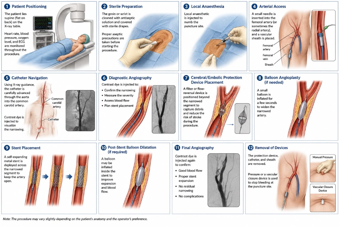
Step 1: Patient Positioning
-The patient lies supine (flat on back) on the X-ray table. Heart rate, blood pressure, oxygen level, and ECG are monitored throughout the procedure.
Step 2: Sterile Preparation
-The groin or wrist is cleaned with antiseptic solution and covered with sterile drapes. Proper aseptic precautions are taken before starting the procedure.
Step 3: Local Anaesthesia
-Local anaesthetic is injected to numb the puncture site.
Step 4: Arterial Access
-A small needle is inserted into the femoral artery (or sometimes the radial artery), and a vascular sheath is placed.
Step 5: Catheter Navigation
-Using X-ray guidance, the catheter is carefully advanced through the aorta into the common carotid artery.
-Contrast dye is injected to visualize the narrowing.
Step 6: Diagnostic Angiography
-Contrast dye is injected to:
- Confirm the narrowing
- Measure the severity
- Assess blood flow
- Plan stent placement
Step 7: Cerebral/ Embolic Protection Device Placement
-A filter or flow-reversal device is positioned beyond the narrowed segment to capture debris and reduce the risk of stroke during the procedure.
Step 8: Balloon Angioplasty (if needed)
-A small balloon is inflated for a few seconds to widen the narrowed artery.
Step 9: Stent Placement
-A self-expanding metal stent is deployed across the narrowed segment to keep the artery open.
Step 10: Post-Stent Balloon Dilatation (if required)
-A balloon may be inflated inside the stent to improve expansion and blood flow.
Step 11: Final Angiography
-Contrast dye is injected again to confirm:
- Good blood flow
- Proper stent expansion
- No residual narrowing
- No complications
Step 12: Removal of Devices
-The protection device, catheter, and sheath are removed.
-Pressure or a vascular closure device is used to stop bleeding at the puncture site.
Duration of the procedure:
-The procedure usually takes 1–2 hours, depending on the complexity and patient condition.
Post-Procedure Care
- Monitor blood pressure, pulse, and neurological status.
- Keep the puncture site clean and dry, asses bleeding or hematoma
- Drink plenty of fluids (unless restricted) to help flush out the contrast.
- Continue antiplatelet medications as prescribed.
- Avoid heavy lifting and strenuous activity for several days.
- Attend follow-up appointments and imaging studies.
Recovery
- Most patients stay in the hospital for 1 day.
- Normal daily activities are usually resumed within a few days, depending on the doctor’s advice.
Follow-Up
-Regular follow-up may include:
- Clinical examination
- Carotid Doppler ultrasound
- Blood pressure and cholesterol management
- Lifestyle modification
- Long-term antiplatelet therapy, if prescribed
-Lifestyle Changes to Prevent Further Disease
- Stop smoking
- Control blood pressure
- Control diabetes
- Lower cholesterol
- Exercise regularly, at least daily graded walking for 30 minutes.
- Maintain a healthy weight
- Eat a heart-healthy diet rich in fruits, vegetables, whole grains, and lean proteins
- Take medications as prescribed
Possible Risks and Complications
-Although generally safe, possible complications include:
- Stroke or TIA
- Bleeding or hematoma at the puncture site
- Artery injury or dissection
- Blood clot formation
- Embolization of plaque debris
- Contrast allergy
- Kidney injury from contrast
- Low heart rate or low blood pressure during balloon inflation
- Restenosis (re-narrowing)
- Infection (rare)
Benefits
- Minimally invasive
- No large neck incision, pin hole procedure.
- Shorter recovery than surgery
- Improves blood flow to the brain
- Reduces the risk of future stroke
- Suitable for many patients at high surgical risk
Would you like to request an appointment?
You can call on +91-98200 86520 for Appointments or fill the form below
If you’re experiencing symptoms of brain vascular issues, contact us today to schedule a consultation and explore your treatment options. Early diagnosis can prevent serious complications and improve your health outcomes.