
Brain Arteriovenous Malformation Embolization
- Home
- Brain Arteriovenous Malformation Embolization
What is an Arteriovenous Malformation (AVM)?
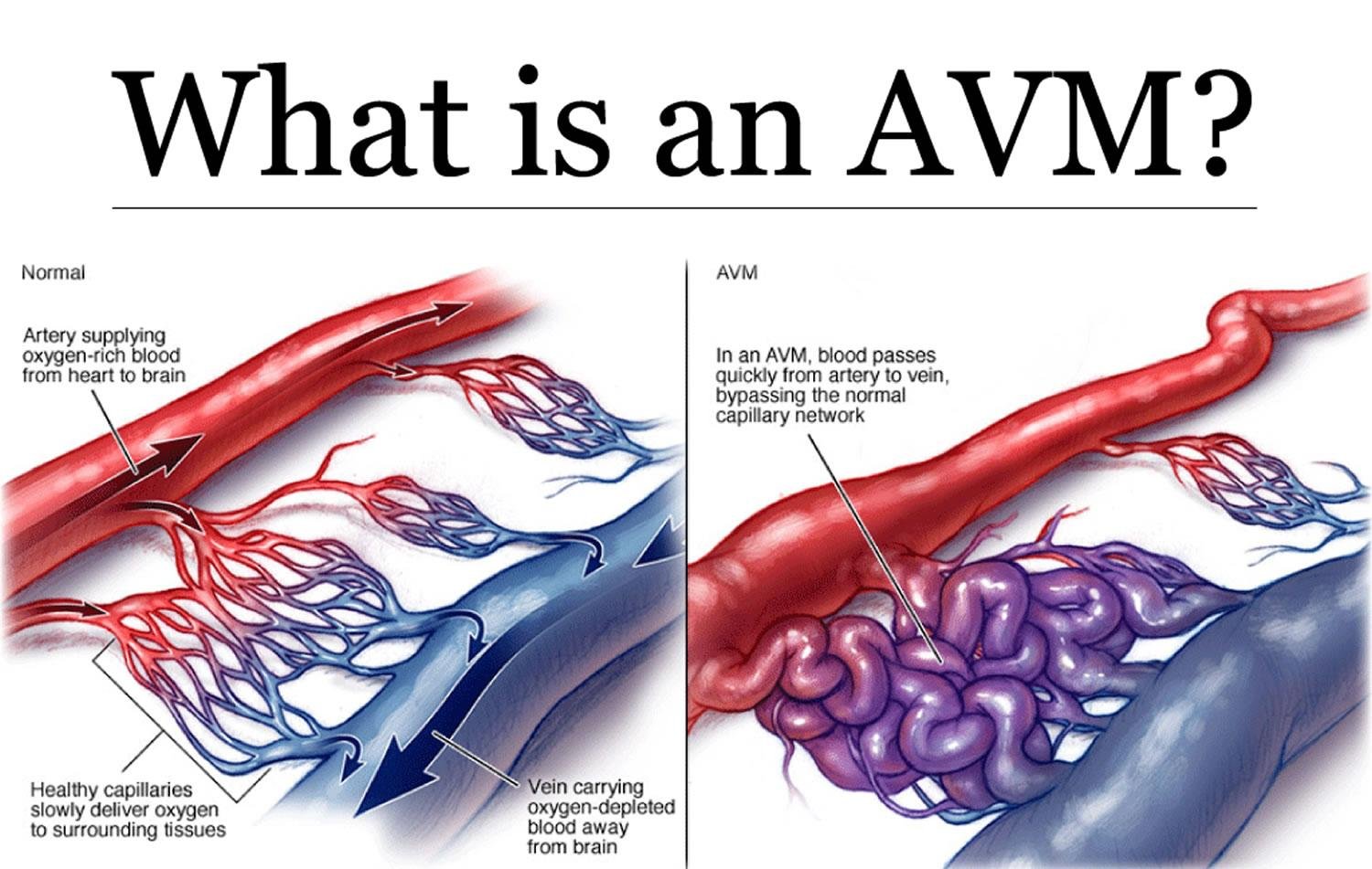
Arteriovenous malformation (AVM) is an abnormal tangle of blood vessels where arteries connect directly to veins without normal capillaries in between.
Normally:
- Arteries carry oxygen-rich blood from the heart.
- Capillaries slow down blood flow and supply oxygen to tissues.
- Veins carry oxygen-poor blood back to the heart.
-In an AVM, blood flows directly from arteries into veins at high pressure, which can damage blood vessels and increase the risk rupture and can cause bleeding.
Where can AVMs occur?
-AVMs can develop almost anywhere in the body, including:
- Brain (most common and most dangerous)
- Spinal cord
- Face and scalp
- Neck
- Arms and legs
- Lungs
- Liver
- Kidneys
- Pelvis
Let us in detail know specifically about Cerebral/ Brain Arteriovenous Malformation:
What is a Brain AVM?
-Brain arteriovenous malformation (Brain AVM) is an abnormal tangle of blood vessels in the brain where arteries connect directly to veins without the normal capillary network.
-In a brain AVM, blood flows directly from arteries into veins under high pressure. This causes the veins to enlarge and weakens their walls, increasing the risk of rupture and bleeding.
Anatomy of a Brain AVM
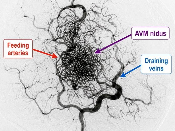
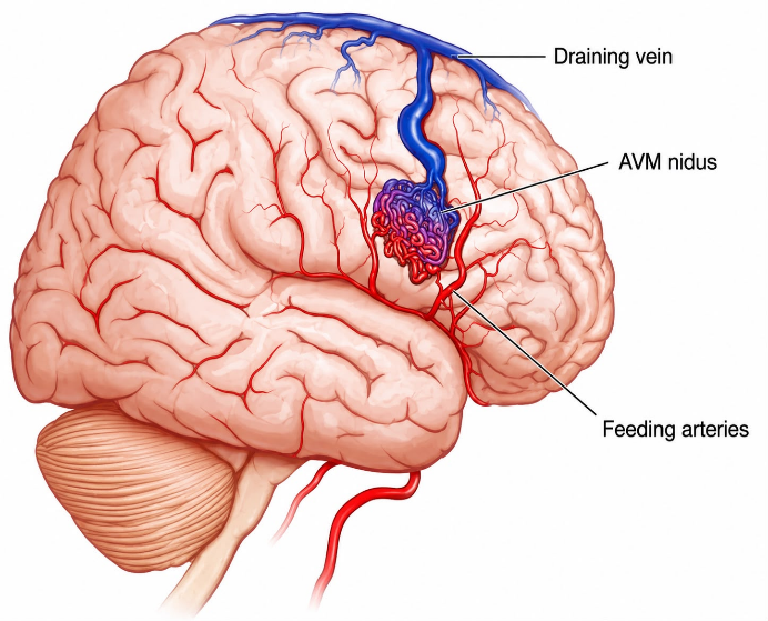
A brain AVM has three main parts:
- Feeding Arteries-Arteries that carry high-pressure blood into the AVM.
- Nidus-The central tangled network of abnormal blood vessels.
- Draining Veins– Veins that carry blood away from the AVM.
How Does a Brain AVM Develop?
Most brain AVMs are congenital, meaning they develop before birth during the formation of blood vessels.
The exact cause is unknown, most causes are:
- Not inherited
- Not caused by lifestyle
- Present at birth, although symptoms may appear later in life
Where Can Brain AVMs Occur?
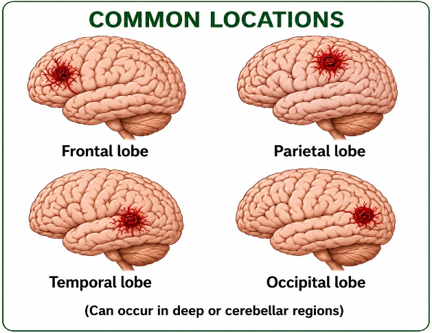
They can develop in any part of the brain, including:
- Frontal lobe
- Parietal lobe
- Temporal lobe
- Occipital lobe
- Cerebellum
- Brainstem (less common but more challenging to treat)
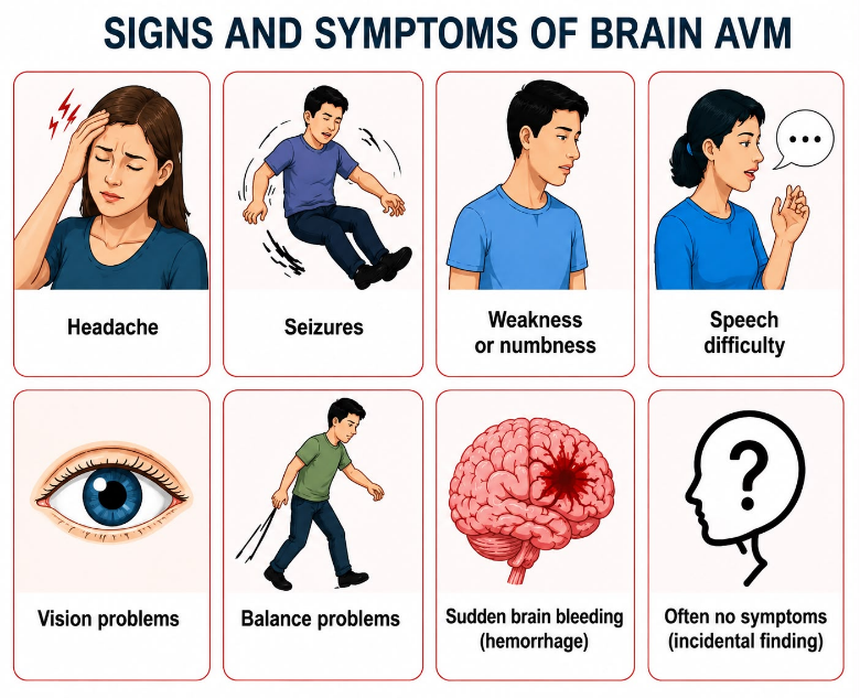
Symptoms
-Some people have no symptoms until the AVM is discovered during imaging or after bleeding.
Common symptoms include:
- Sudden severe headache
- Seizures
- Weakness of the face, arm, or leg
- Numbness
- Difficulty speaking
- Vision problems
- Loss of balance
- Dizziness
- Memory problems
- Ringing sound in the head (rare)
- Brain haemorrhage
What Happens if an AVM Ruptures?
A ruptured AVM can cause bleeding into or around the brain.
-Symptoms include:
- Sudden “worst headache of life”
- Vomiting
- Loss of consciousness
- Seizures
- Paralysis
- Difficulty speaking
- Vision loss
- Confusion
- Coma (severe cases)
This is a medical emergency requiring immediate treatment.
Risk Factors for Rupture
- Congenital vascular abnormality
- Previous AVM bleed
- Pregnancy may increase blood flow but has not been proven to significantly increase rupture risk in all patients
Complications for Rupture
If untreated, complications may include:
- Brain haemorrhage
- Stroke
- Permanent neurological deficits
- Seizures
- Hydrocephalus (fluid buildup in the brain)
- Brain damage
- Death (in severe haemorrhage)
Diagnosis
1. Medical History
The doctor asks about- Headaches, seizures, previous bleeding weakness, vision changes, speech problems, family history, current medications and allergies.
2. Physical Examination
A neurological examination includes- Consciousness, speech, vision, cranial nerves, muscle strength, sensation, coordination, reflexes and balance.
3. Blood Tests
- Complete blood count (CBC)
- Kidney function tests
- Liver function tests
- PT/INR
- aPTT
- Blood sugar
- Blood group and cross-match (if needed)
4. Imaging Tests
(a) CT Scan– Used in emergencies to detect:
- Brain hemorrhage
- Swelling
- Large AVMs
(b) MRI Brain– Shows:
- Exact location
- Size
- Relationship to surrounding brain tissue
- Previous bleeding
(c) MR Angiography (MRA)- Shows:
- Feeding arteries
- Nidus
- Draining veins
- Blood flow pattern
(d) CT Angiography (CTA)- Provides detailed images of blood vessels and helps with treatment planning.
(e) Digital Subtraction Angiography (DSA)
-This is the gold standard investigation for brain AVMs, it shows:
- Feeding arteries
- Nidus
- Draining veins
- Blood flow dynamics
- Exact anatomy for treatment planning
Treatment Options for Brain AVM
-Treatment depends on:
- Age
- Symptoms
- AVM size
- Location
- Risk of bleeding
- Overall health
Options include:
- Observation– For selected low-risk AVMs.
- Microsurgical Removal– Open surgery to completely remove the AVM.
- Stereotactic Radiosurgery– Focused radiation gradually closes the AVM over months to years.
- Endovascular Embolization– Blocks the abnormal blood vessels using a catheter.
-Sometimes, a combination of these treatments is used.
–Let us know in detail about Cerebral/ Brain Arteriovenous Malformation Embolization:
What is Brain AVM Embolization?
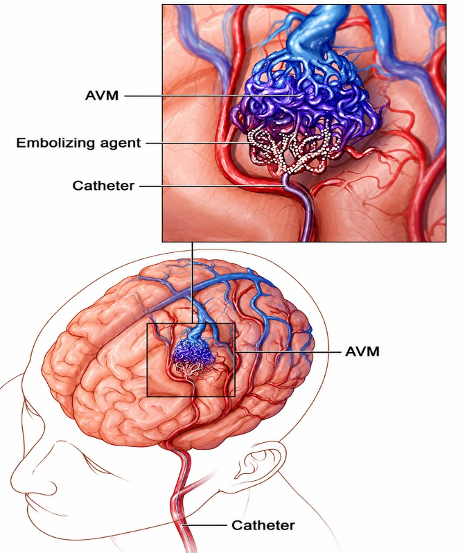
-Brain AVM embolization is a minimally invasive endovascular procedure performed by an interventional neuroradiologist or endovascular neurosurgeon.
-A tiny catheter is guided through an artery (usually in the wrist or groin) into the arteries supplying the AVM. A special liquid or other embolic material is injected to block the abnormal blood vessels.
The goal is to:
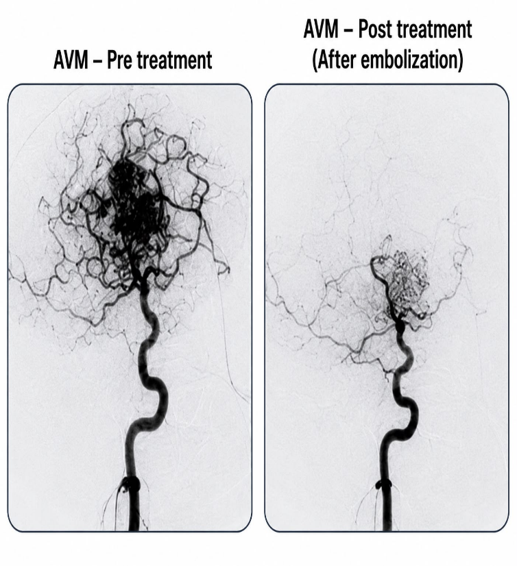
- Reduce blood flow through the AVM.
- Decrease the risk of bleeding.
- Reduce AVM size and reduce symptoms.
- Treat ruptured AVMs in selected Situations.
- Prepare the AVM for surgery or radiosurgery and improve its effectiveness.
Who Needs Brain AVM Embolization?
-It may be recommended for patients with:
- Brain AVM discovered on imaging.
- Previous brain haemorrhage.
- Seizures caused by an AVM.
- Severe headaches related to the AVM.
- Progressive neurological symptoms.
- High-risk AVMs with weak areas or aneurysms.
- AVMs planned for surgery or radiosurgery.
When is Embolization NOT Recommended?
-It may not be suitable if:
- The AVM is very small and better treated by surgery or radiosurgery alone.
- The AVM is located in an area where embolization carries excessive risk.
- The feeding arteries cannot be safely reached.
- The patient’s overall health makes the procedure too risky.
Preparation Before Embolization
Patients are usually advised to:
- Fast for several hours before the procedure.
- Review medications with the doctor.
- Stop certain blood thinners if instructed.
- Sign an informed consent form.
- Change into a hospital gown.
- Have an IV line placed.
Anaesthesia
-Usually performed under general anaesthesia, although selected cases may use conscious sedation.
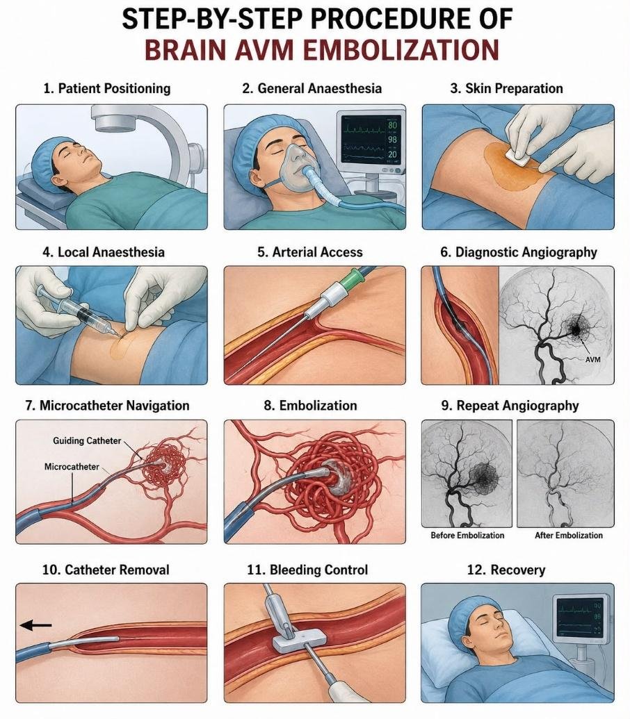
Step by step procedure
- The patient lies supine (flat on back) on the angiography table.
- Heart rate, blood pressure, oxygen level, and ECG are continuously monitored.
General anaesthesia is given so the patient remains still and comfortable throughout the procedure.
The selected access site, usually the wrist or groin (radial or femoral artery puncture), is cleaned with an antiseptic solution and covered with sterile drapes.
Local anaesthetic is injected at the arterial puncture site to numb the area.
The radial or femoral artery is punctured, and a small vascular sheath is inserted to provide access for the catheters.
Contrast dye is injected, and X-ray images are taken to identify the AVM, its feeding arteries, nidus, and draining veins.
A very thin microcatheter is carefully guided through the blood vessels toward the arteries supplying the AVM.
The selected embolic material is injected through the microcatheter to block abnormal blood flow within the AVM while preserving normal brain vessels.
Further angiographic images are obtained to assess the degree of AVM occlusion and check for residual abnormal blood flow.
The microcatheter, guiding catheter, and vascular sheath are carefully removed after the treatment is completed.
The arterial puncture site is closed using manual pressure or a vascular closure device to prevent bleeding.
The patient is shifted to a recovery area or ICU for neurological and vital-sign monitoring after the procedure.
After the Procedure
-Patients are monitored for:
- Blood pressure
- Heart rate
- Oxygen levels
- Neurological status
- Access site bleeding
- Headache
- Signs of stroke or hemorrhage
Recovery at Home
-Patients are generally advised to:
- Rest for several days.
- Avoid heavy lifting for about a week or as instructed.
- Drink plenty of fluids unless restricted.
- Take prescribed medications.
- Keep the puncture site clean and dry.
- Attend scheduled follow-up appointments.
Benefits
- Minimally invasive (no open brain surgery incision)
- Reduces blood flow to the AVM
- Lowers the risk of bleeding in selected patients
- Can shrink the AVM
- Makes surgery safer
- Improves radiosurgery outcomes
- May completely cure some small AVMs
Risks and Complications
-Although generally safe in experienced hands, possible complications include:
- Stroke
- Brain hemorrhage
- Temporary or permanent neurological deficits
- Seizures
- Vessel injury
- Blood clot formation
- Brain swelling
- Contrast allergy
- Kidney injury from contrast (rare)
- Infection
- Groin or wrist hematoma
- Failure to completely close the AVM
- Need for repeat embolization
Follow-Up
-Follow-up may include:
- Neurological examination
- MRI/MRA
- Cerebral angiography
- Additional embolization sessions if needed
- Surgery or radiosurgery when planned
Warning Signs After Discharge
-Seek emergency medical care immediately if you develop:
- Sudden severe headache
- Weakness or numbness
- Difficulty speaking
- Vision loss
- Confusion
- Seizures
- Persistent vomiting
- Loss of consciousness
- Severe bleeding from the access site
Frequently asked questions (FAQs)
1. Is brain AVM embolization a surgery?
-No. It is a minimally invasive catheter-based procedure performed through a blood vessel.
2. Is the procedure painful?
-No. It is usually done under general anaesthesia, so you do not feel pain during the procedure.
3. Can embolization cure a brain AVM?
-Some small AVMs can be completely cured, but many require additional surgery or radiosurgery.
4. Will I need more than one embolization session?
-Yes. Large or complex AVMs often require staged embolization over multiple sessions.
5. Can an AVM come back after successful treatment?
-Recurrence is uncommon in adults after complete obliteration but can occur, particularly in children, so follow-up imaging is important.
6. Is embolization always the first treatment?
-Not always. The treatment plan depends on the AVM’s size, location, bleeding risk, and the patient’s overall health. Sometimes surgery or radiosurgery is preferred, and embolization is used as an adjunct.
Would you like to request an appointment?
You can call on +91-98200 86520 for Appointments or fill the form below