
Pelvic Venous Congestion Syndrome Embolization
- Home
- Pelvic Venous Congestion Syndrome Embolization
Pelvic venous congestion syndrome embolization is a minimally invasive procedure performed by an interventional radiologist to block the abnormal pelvic veins causing congestion.
-Tiny coils, plugs, or a special liquid/sclerosant are placed inside the affected veins to stop the abnormal blood flow. Blood is naturally redirected through healthy veins, relieving pressure and reducing pain.
How does embolization work?
-Instead of removing the veins, embolization blocks the faulty veins, allowing blood to flow through healthy veins. Over time, the blocked veins shrink, reducing pressure and relieving pain.
What is Pelvic Venous Congestion Syndrome (PVCS)?
-Pelvic Venous Congestion Syndrome (PVCS), also called Pelvic Congestion Syndrome (PCS), is a condition in which the veins in the pelvis become enlarged, stretched, and unable to carry blood efficiently. This causes blood to pool in the pelvic veins, leading to chronic pelvic pain.
-It is similar to varicose veins in the legs, but it occurs inside the pelvis.
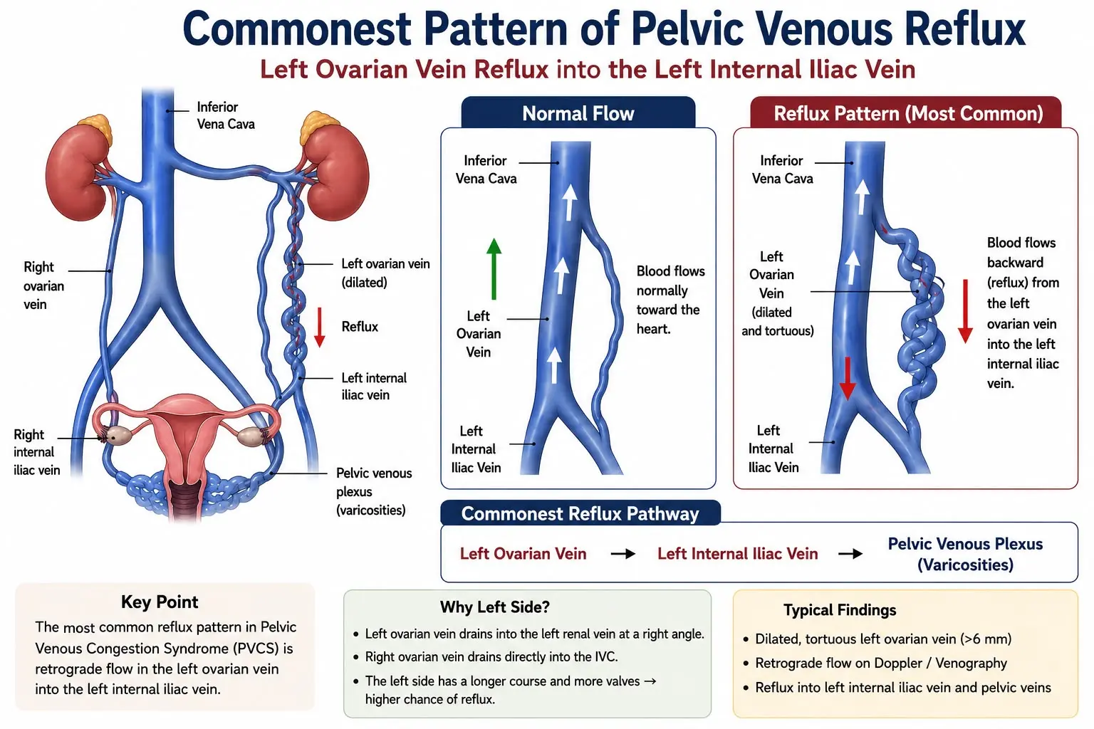
-Normally, veins contain one-way valves that prevent blood from flowing backward. When these valves become weak or damaged, blood flows backward (called venous reflux) and collects in the pelvic veins. This causes the veins to become dilated (varicose veins), leading to chronic pelvic pain.
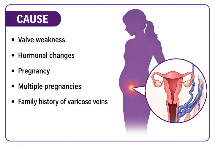
Causes of PVCS:
- Pregnancy (most common)
- Multiple pregnancies
- Weak or damaged vein valves
- Hormonal changes (especially estrogen)
- Enlarged ovarian veins
- Compression of pelvic veins
- Family history of varicose veins
Risk Factors of PVCS:
- Women aged 20–45 years
- Multiple pregnancies
- Standing for long periods
- Hormonal changes
- Varicose veins in the legs
- Family history
Symptoms of PVCS:
Your doctor may recommend embolization if:
- Symptoms are severe.
- Medicines have not helped.
- Imaging confirms enlarged pelvic veins.
- The pain affects daily activities.
Pre-Procedure Evaluation:
A. Clinical Assessment
- Duration of pelvic pain
- Menstrual history
- Previous pregnancies- commonest cause of PVCS
- Pain during intercourse
- Previous pelvic surgery
- Varicose veins
B. Physical Examination
- Pelvic tenderness
- Vulvar varicose veins
- Leg varicose veins
C. Blood Tests
- Complete Blood Count (CBC) : for anemia and trace of any infection
- Kidney function tests : to ensure if it is safe to use contrast dye
- Blood clotting profile (PT/INR) : to know the risk of bleeding
- Pregnancy test (if required)
D. Imaging Tests
1. Pelvic Ultrasound (with Doppler)
Shows:
- Enlarged ovarian veins
- Slow blood flow
- Venous reflux
2. CT Venography
Shows:
- Dilated pelvic veins
- Anatomy of pelvic vessels
- Compression of veins
3. MRI/MR Venography
Shows:
- Pelvic veins clearly
- Other causes of pelvic pain
4. Diagnostic Venography (Gold Standard)
-This is performed immediately before embolization.
It shows:
- Abnormal veins
- Backward blood flow
- Exact veins needing treatment
Preparation Before the Procedure:
The patient may be advised to:
- Fast for a few hours (if sedation is planned)
- Take prescribed antibiotics if advised
- Stop blood thinners if taking 3 days prior
- Empty the bladder
- Avoid intercourse before the procedure if instructed
- Sign informed consent
Where is the procedure performed?
-It is performed in an Interventional Radiology (IR) suite using continuous X-ray (fluoroscopy).
Procedure of pelvic venous congestion syndrome embolization:
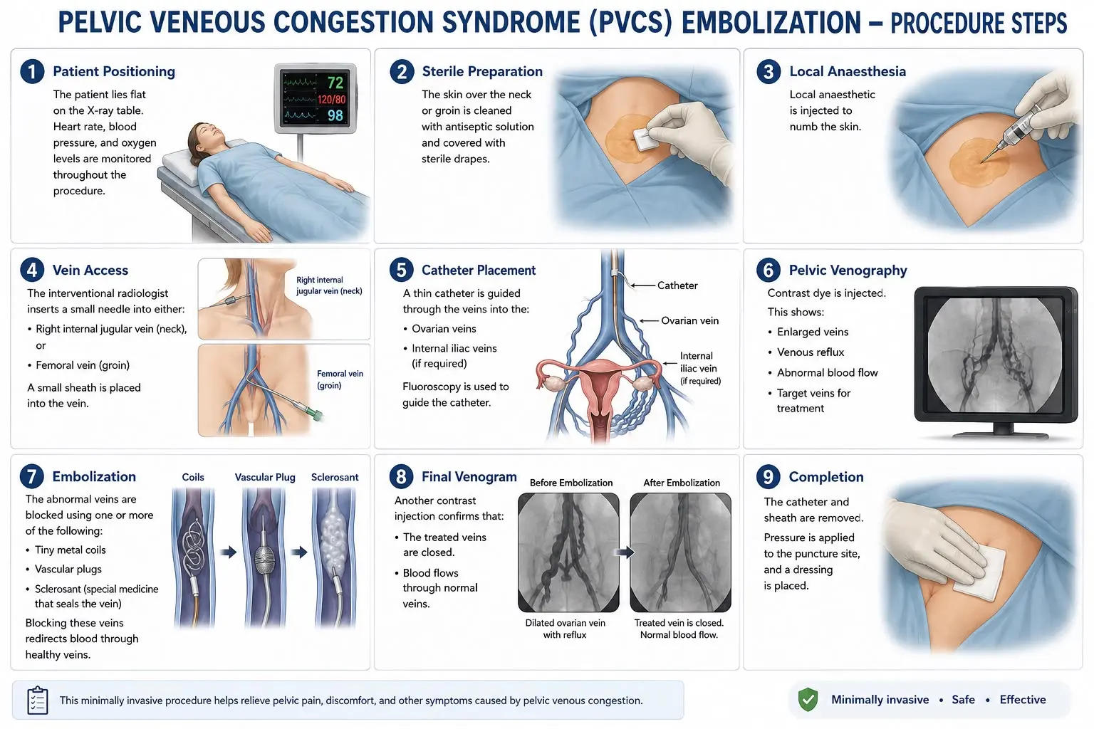
Step 1: Patient Positioning
The patient lies supine (flat on back) on the X-ray table. Heart rate, blood pressure, ECG and oxygen levels are monitored throughout the procedure.
Step 2: Sterile Preparation
The skin over the neck or groin is cleaned with antiseptic solution and covered with sterile drapes.
Step 3: Local Anaesthesia
-Local anaesthetic is injected to numb the skin over the puncture site.
Step 4: Vein Access
-The interventional radiologist inserts a small needle into either:
- Right internal jugular vein (neck), or
- Femoral vein (groin)
-A small sheath is placed into the vein.
Step 5: Catheter Placement/ Navigation
-A thin catheter is guided through the veins into the:
- Ovarian veins
- Internal iliac veins (if required)
Fluoroscopy is used to guide the catheter.
Step 6: Pelvic Venography
-Contrast dye is injected.
-This shows:
- Enlarged veins
- Venous reflux
- Abnormal blood flow
- Target veins for treatment
Step 7: Embolization
-The abnormal veins are blocked using one or more of the following:
- Tiny metal coils
- Vascular plugs
- Sclerosant (special medicine that seals the vein)
Blocking these veins redirects blood through healthy veins.
Step 8: Final/ Post Embolization Venogram
Another contrast injection confirms that:
- The treated veins are closed.
- Blood flows through normal veins.
After procedure care:
Most patients:
- Rest for a few hours
- Go home the same day
- Resume light activities within 24 hours
Recovery:
- Mild pelvic pain or cramping for a few days
- Mild bruising at the puncture site
- Resume light activities in 24–48 hours
- Avoid heavy lifting for about one week
- Drink plenty of fluids to help flush out the contrast dye
Possible Risks:
Although uncommon, risks include:
- Bruising or bleeding at the puncture site
- Infection
- Allergy to contrast dye
- Temporary pelvic pain after embolization
- Coil migration (rare)
- Damage to nearby blood vessels (rare)
- Recurrence of symptoms
Follow-up:
A follow-up visit is usually scheduled within 4–12 weeks. Your doctor may assess symptom improvement and, if needed, perform an ultrasound or other imaging to evaluate the treated veins.
Benefits:
- Minimally invasive (no large incision)
- Preserves the uterus and ovaries
- Quick recovery
- High success rate for pain relief
- Same-day discharge in most cases
- Less pain than open surgery
Would you like to request an appointment?
You can call on +91-98200 86520 for Appointments or fill the form below