
Renal Tumour RFA (Radiofrequency Ablation)
- Home
- Renal Tumour RFA (Radiofrequency Ablation)
Renal Tumour Radiofrequency Ablation (RFA) is a minimally invasive procedure used to destroy a small kidney tumour without removing the kidney.
A thin needle (called an ablation probe) is inserted into the tumour under imaging guidance. The probe delivers high-frequency electrical energy, producing heat (about 60–100°C) that destroys the cancer cells.
RFA is usually performed by an interventional radiologist using CT scan or ultrasound guidance.
What is a Renal Tumour?
– A renal tumour is an abnormal growth in the kidney.
It can be:
- Benign (non-cancerous)
- Malignant (kidney cancer)
-RFA is mainly used for small kidney cancers.
How Does RFA Work?
Radiofrequency energy passes through the probe.
The heat:
- Kills tumour cells
- Stops blood supply to the tumour
- Causes permanent destruction of the tumour tissue
-Over time, the treated tumour shrinks and becomes scar tissue.
Who Needs Renal Tumour RFA?
RFA is recommended for patients with/ suitable candidates:
- Small kidney tumours (usually less than 3–4 cm)
- Early-stage kidney cancer
- Poor surgical fitness
- Elderly patients
- Patients with only one functioning kidney
- Multiple kidney tumours
- Patients wishing to avoid surgery when appropriate
When is RFA Not Suitable?
RFA may not be suitable if:
- Tumour is very large
- Tumour is very close to the collecting system or major blood vessels
- Cancer has spread to other organs
- Uncorrected bleeding disorders
- Active infection
Pre-Procedure Evaluation:
-Before performing Renal Tumour Radiofrequency Ablation (RFA), a thorough evaluation is done to ensure that the procedure is safe and appropriate for the patient.
1. Clinical Assessment-
The doctor reviews the patient’s medical history, previous kidney disease or surgeries, current medications, allergies, symptoms, blood pressure, and overall health to ensure the procedure is safe.
2. Blood Tests
- Complete Blood Count (CBC)- checks levels of RBCs, WBCs and platelet.
- Kidney function tests- Serum Creatinine, blood urea to asses how well the kidneys are working.
- Electrolytes Level- to detect any imbalance.
- PT/INR, aPTT- to ensure normal blood clotting and to reduce the risk of bleeding during the procedure if needed.
- Blood sugar- specially in patients with diabetes.
3. Imaging-
Imaging helps determine the tumour’s size and location.
-Common studies include:
- Ultrasound
- Contrast-enhanced CT scan
- MRI (if needed)
4. Consent–
The doctor explains the procedure, its benefits, risks, alternatives, and possible complications. Written informed consent is obtained before the procedure.
Patient Preparation:
Before the procedure:
- Fast for about 6–8 hours
- Stop blood thinners if advised
- Wear a hospital gown
- An IV line is inserted
- Vital signs are monitored
- Antibiotics may be given
- Sedation or general anaesthesia is planned
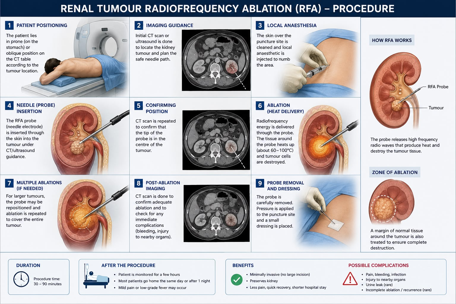
Procedure of Renal Tumour Radiofrequency Ablation (RFA):
-The patient is positioned on the CT scan table, usually lying on the stomach or side, depending on the location of the kidney tumour.
-A CT scan or ultrasound is performed to accurately locate the tumour and plan the safest path for inserting the RFA needle.
-The skin over the puncture site is cleaned with an antiseptic solution, and a local anaesthetic is injected to numb the area. Sedation or general anaesthesia may also be given if required.
-A thin RFA probe is carefully inserted through the skin into the centre of the kidney tumour under continuous imaging guidance.
-A repeat CT scan or ultrasound is performed to confirm that the tip of the probe is correctly positioned within the tumour before treatment begins.
-Radiofrequency energy is delivered through the probe, generating heat of about 60–100°C to destroy the tumour cells while preserving as much healthy kidney tissue as possible.
-For larger tumours, the probe may be repositioned and additional ablations performed to ensure the entire tumour is treated.
-A final CT scan or ultrasound is performed to confirm that the tumour has been adequately treated and to check for any immediate complications.
-The RFA probe is carefully removed, pressure is applied to the puncture site to prevent -bleeding, and a small sterile dressing is placed over the skin.
After the Procedure:
–Patients are monitored for:
- Pain
- Bleeding
- Blood pressure
- Urine output
- Fever
-Most patients go home the same day or after one night’s stay.
Recovery
Most people:
- Walk within a few hours
- Resume light activities in 1–3 days
- Return to normal activities in about 1 week
-Heavy lifting should be avoided for 1–2 weeks
Follow-Up:
-Follow-up imaging is essential to ensure the tumour has ben completely treated and checks for recurrence.
Usually:
- CT scan or MRI at 1–3 months
- Then every 6–12 months as advised
- Blood tests to asses kidney functioning
Possible Risks and Complications:
Most complications are uncommon, but may include:
- Pain
- Bleeding
- Bruising
- Infection
- Injury to nearby organs
- Damage to the urine collecting system
- Urine leakage (rare)
- Temporary blood in urine
- Skin burn (very rare)
- Incomplete tumour destruction
- Tumour recurrence
Major complications are uncommon.
Advantages of RFA:
- No large surgical incision
- Kidney is preserved
- Less pain
- Short hospital stay
- Faster recovery
- Lower risk of complications
- Can be repeated if needed
- Suitable for high-risk surgical patients
Limitations:
- Not suitable for all tumours
- Larger tumours may not be completely treated
- Rare need for repeat ablation
- Requires follow-up imaging
Would you like to request an appointment?
You can call on +91-98200 86520 for Appointments or fill the form below