
Post Transplant Renal Artery Stenosis Angioplasty / Stenting
- Home
- Post Transplant Renal Artery Stenosis Angioplasty / Stenting
Post-transplant renal artery stenosis (TRAS) is the narrowing of the renal artery supplying a transplanted kidney. It is one of the most common vascular complications after kidney transplantation and is a potentially reversible cause of:
- Resistant hypertension
- Worsening kidney function
- Fluid overload
- Graft dysfunction
-The narrowing reduces blood flow to the transplanted kidney, activating the renin-angiotensin-aldosterone system (RAAS), leading to hypertension and impaired kidney function.
-Percutaneous Transluminal Angioplasty (PTA), with or without stent placement, is considered the first-line treatment for significant TRAS.
Anatomy
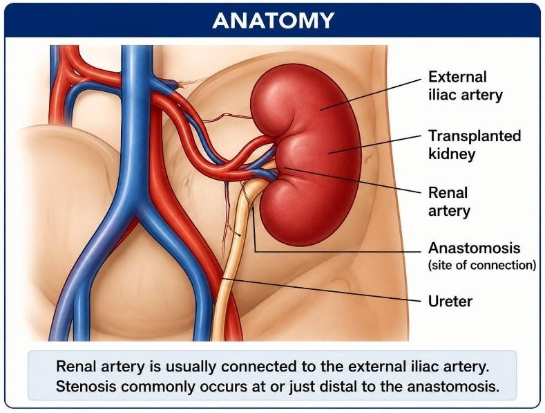
The transplanted kidney is usually placed in the:
- Right iliac fossa (most common)
- Left iliac fossa (occasionally)
The renal artery is connected to:
- External iliac artery
- Internal iliac artery
- Common iliac artery
TRAS usually occurs:
- At the arterial anastomosis (most common)
- Just distal to the anastomosis
- Within the donor renal artery
- Rarely within the recipient iliac artery
Causes:
Surgical Causes
- Anastomotic scarring– The anastomosis is the site where the donor renal artery is surgically connected to the recipient’s iliac artery. During healing, excessive scar tissue can form at this junction, narrowing the artery and reducing blood flow to the transplanted kidney.
- Technical problems during surgery– If the artery is not connected properly during the transplant (for example, due to slight narrowing, twisting, or tension at the anastomosis), blood flow may be restricted, leading to stenosis over time.
- Kinking of the artery– If the transplanted renal artery is too long or is positioned awkwardly, it may bend or kink. This decreases blood flow to the kidney.
- Vessel torsion– Rarely, the transplanted kidney or its artery may rotate after surgery, causing the artery to twist. This can significantly reduce or even block blood flow and requires urgent treatment.
Donor Factors
These are conditions related to the donated kidney or donor artery.
- Atherosclerosis- Older donors or donors with vascular disease may have atherosclerosis (fatty plaque buildup) in the renal artery. This plaque can cause narrowing before or after transplantation.
- Small-caliber artery- Some donor kidneys have naturally small renal arteries, making the surgical connection more difficult and increasing the risk of postoperative narrowing.
- Multiple renal arteries- Some kidneys have two or more renal arteries instead of one. Reconstructing multiple arteries is technically more complex and increases the risk of stenosis at the anastomosis.
Recipient Factors
These are conditions present in the kidney transplant recipient.
- Iliac artery disease– The donor renal artery is connected to the recipient’s iliac artery. If the recipient has narrowing or plaque in the iliac artery, blood flow to the transplanted kidney may be reduced.
- Diabetes– Diabetes damages blood vessels by accelerating atherosclerosis and promoting inflammation.
- Hypertension– Long-standing high blood pressure damages the arterial wall, making blood vessels stiffer and more prone to narrowing.
- Smoking– Smoking injures the lining of blood vessels (endothelium), promotes plaque formation, and accelerates vascular disease.
- Hyperlipidemia– High cholesterol levels contribute to plaque buildup in arteries, which can narrow the renal or iliac arteries over time.
Other Causes
- Fibrosis– Excessive healing after surgery may produce fibrous (scar) tissue around the artery. This tissue can compress the vessel externally or narrow it from within.
- Intimal hyperplasia– The intima is the innermost layer of the artery. Injury from surgery or angioplasty can stimulate smooth muscle cells to proliferate, causing thickening of this layer and progressive narrowing of the artery.
- Rejection causing vascular injury– During acute or chronic rejection, the body’s immune system attacks the transplanted kidney, including its blood vessels. This inflammation damages the artery and can lead to stenosis.
- External compression– Structures such as a hematoma (blood collection), lymphocele (collection of lymphatic fluid), postoperative swelling, or nearby scar tissue may press on the renal artery from outside, reducing blood flow.
- Infection (rare)- Severe infections involving the transplanted kidney or surrounding tissues can inflame and weaken the artery, leading to narrowing or, rarely, aneurysm formation.
Risk Factors:
- Older donor age
- Delayed graft function
- Multiple renal arteries
- Difficult surgery
- Previous vascular disease
- Diabetes
- Smoking
- Hyperlipidemia
Symptoms:
Many patients are initially asymptomatic.
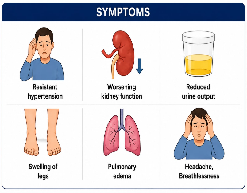
-Common symptoms include:
- Resistant hypertension
- Worsening kidney function
- Reduced urine output
- Pulmonary edema
- Headache
- Breathlessness
- Fluid overload
Clinical Signs:
- High blood pressure
- Bruit over transplanted kidney
- Leg edema
- Elevated creatinine
- Reduced estimated GFR
Diagnosis:
1. Laboratory Tests-
- Serum creatinine, blood urea nitrogen (BUN), eGFR, serum electrolytes, complete blood count, coagulation profile (PT/INR).-
2. Imaging
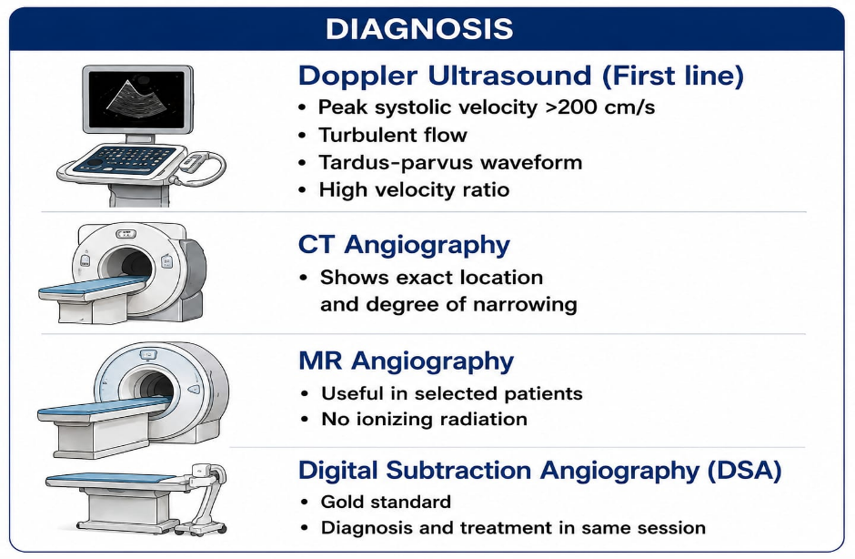
- Doppler Ultrasound– First-line investigation.
- CT Angiography
Shows:
-Exact location
– Degree of narrowing
– Vascular anatomy
MR Angiography:
– Useful in selected patients.
Digital Subtraction Angiography (DSA) :
– Gold standard.
Allows:
– Diagnosis
– Immediate angioplasty
– Stent placement
Indications for Angioplasty/Stenting:
-Treatment is recommended when significant stenosis causes:
- Resistant hypertension
- Rising serum creatinine
- Declining graft function
- Pulmonary edema
- Significant angiographic narrowing (>50–70%) with pressure gradient
- Symptomatic renal artery stenosis
Contraindications:
Absolute
- Active uncontrolled infection
- Severe contrast allergy (unless managed appropriately)
- Patient refusal
Relative
- Coagulopathy (bleeding disorder)
- Severe thrombocytopenia (low platelet count)
- Uncontrolled hypertension
- Pregnancy (rare)
- Difficult vascular anatomy
Pre-Procedure Evaluation:
- Clinical Assessment– The doctor reviews the patient’s medical history, kidney transplant details, current medications, blood pressure, fluid status, and urine output.
- Blood Test– including CBC, serum creatinine, blood urea, electrolytes, PT/INR, aPTT, platelet count, and blood sugar, are performed to assess kidney function and ensure it is safe to proceed.
- Imaging Review- Previous Doppler ultrasound, CT angiography, angiograms, and kidney function trends are reviewed to identify the location and severity of the stenosis.
- Medication Review–The doctor reviews anticoagulants, antiplatelet drugs, immunosuppressive medications, and any history of allergies to plan the procedure safely.
- Consent– The procedure, including its benefits, risks, alternatives, the possible need for a stent, and potential complications, is explained, and informed consent is obtained.
Procedure of Post-Transplant Renal Artery Angioplasty/Stenting:
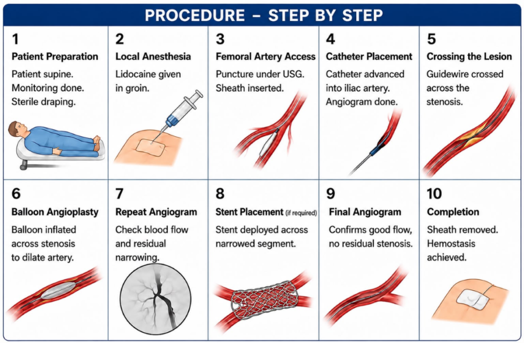
Patient lies supine (flat on the back) on angiography table. The groin is cleaned with antiseptic solution. Sterile drapes are applied. ECG, blood pressure, and oxygen saturation are monitored.
Local anesthesia Lidocaine is injected into the groin skin to numb the puncture site. Conscious sedation may be given if needed.
Under ultrasound guidance, the common femoral artery is punctured and vascular sheath inserted to provide safe pathway for catheter or guide wire.
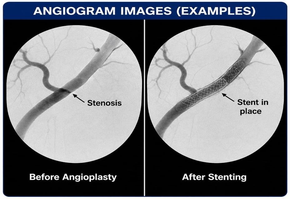
A guide catheter is advanced into iliac artery supplying the transplanted kidney. Contrast dye is injected to get an initial angiogram which identifies:
- Location of stenosis
- Severity
- Vessel anatomy
Soft guidewire is carefully passed across narrowed segment of the renal artery
A Balloon catheter is positioned across stenosis and inflated for several seconds. This Narrows the plaque and expands artery to improve blood flow.
A repeat angiogram is performed to check
- Blood flow
- Residual narrowing
- Vessel injury
- A stent indicated when there is significant residual stenosis, elastic recoil occur, dissection after angioplasty present.
- The stent is deployed across the narrowed segment and expanded with a balloon (if balloon-expandable).
Confirms:
- Excellent blood flow
- No residual stenosis
- Patent artery
- Good graft perfusion
The Catheter and sheath are removed, and bleeding is controlled using manual compression, or a vascular closure device. Sterile dressing applied to punctured skin.
During the Procedure:
Patients are monitored for:
- Blood pressure
- Heart rate
- Oxygen saturation
- Pain
- Contrast reactions
- Kidney perfusion
- Urine output (if applicable)
Post-Procedure Care:
Monitoring-
- Blood pressure
- Pulse
- Oxygen saturation
- Puncture site
- Urine output
- Serum creatinine
Bed Rest-
Usually for 4–6 hours (depending on access site and closure device)
Medications-
- Antiplatelet therapy (e.g., aspirin, sometimes with clopidogrel as advised)
- Continue immunosuppressive medications
- Pain medication if required
- Adequate hydration (when appropriate)
Imaging Follow-Up-
- Doppler ultrasound within 24–48 hours or as advised
- Periodic Doppler surveillance during follow-up
Complications:
Access Site-
- Bleeding
- Hematoma
- Pseud-oaneurysm
- Arteriovenous fistula
Vessel Complications-
- Arterial dissection
- Arterial rupture (rare)
- Thrombosis
- Distal embolization
- Vasospasm
Kidney Complications-
- Contrast-induced kidney injury
- Temporary graft dysfunction
- Rare graft infarction
Stent Complications
- Stent thrombosis
- Restenosis
- Stent migration (rare)
- In-stent restenosis
General Complications
- Contrast allergy
- Infection
- Arrhythmias (rare)
- Death (extremely rare)
Advantages of Angioplasty/Stenting:
- Minimally invasive
- Preserves transplanted kidney
- Improves blood flow
- Improves graft function
- Controls blood pressure
- Short hospital stay
- High technical success (over 90% in experienced centers)
- Faster recovery than surgery
Would you like to request an appointment?
You can call on +91-98200 86520 for Appointments or fill the form below