
Percutaneous Nephrostomy (PCN)
- Home
- Percutaneous Nephrostomy (PCN)
-Percutaneous Nephrostomy (PCN) is an image-guided procedure in which a thin catheter (tube) is inserted through the skin directly into the kidney’s collecting system to drain urine when the normal urinary pathway is blocked or not functioning properly.
-A PCN creates an alternative route for urine drainage from the kidney to an external drainage bag. It helps relieve pressure on the kidney, prevents kidney damage, and treats or prevents infection caused by urinary obstruction
Normally, urine flows from the kidney → ureter → bladder → urethra. When there is an obstruction, urine accumulates in the kidney causing hydronephrosis. PCN bypasses the obstruction and drains urine externally.
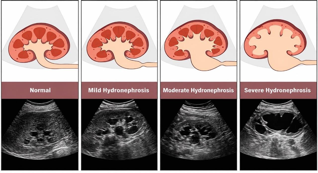
Indications for PCN:
1. Obstructive Causes
- Ureteric stones
- Ureteric strictures
- Pelvi-ureteric junction (PUJ) obstruction
- Bladder outlet obstruction
- Blood clots causing urinary obstruction
2. Malignant Causes
- Cervical/Prostate/ Bladder cancer
- Colorectal cancer causing ureteric compression
- Retroperitoneal tumors
3. Infectious Causes
- Pyonephrosis (infected obstructed kidney)
- Severe urinary tract infection with obstruction
4. Other Indications
- Urinary diversion after trauma
- Urinary leak or fistula
- Access for further urological procedures
- Antegrade ureteric stenting
- Diagnostic nephrostogram
Contraindications:
1. Absolute
- No safe access route to kidney
- Uncorrectable severe bleeding disorder
2. Relative
- Coagulopathy (bleeding disorder)
- Anticoagulant therapy
- Pregnancy (requires special precautions)
- Uncontrolled hypertension
- Severe obesity
Pre-procedure Evaluation:
Clinical Assessment
- History and physical examination
- Assessment of symptoms: Flank pain, fever, reduced urine output, hematuria.
Laboratory Tests
- Complete blood count (CBC), Platelet count.
- Coagulation profile- PT/INR- to assess the risk of hemorrhage.
- Renal function tests
- Serum creatinine
- Urine culture
Imaging- To confirm the indications of the procedure and to asses renal anatomy.
- Ultrasound abdomen
- CT KUB (Kidney, Ureter, Bladder)
- CT urography if needed
Consent- The patient is informed about the procedure, possible risks, benefits and complications to the patient and relatives by the Interventional Radiologist. Written content is also taken.
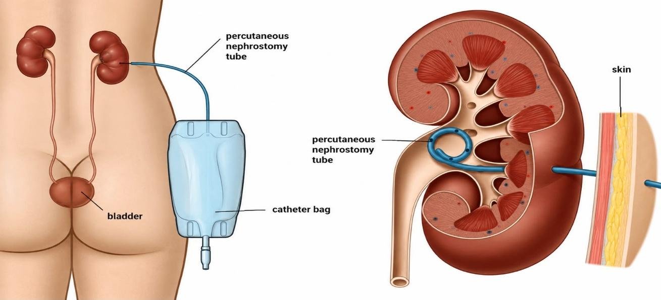
Procedure of Percutaneous Nephrostomy (PCN):
The patient is positioned in the prone or slightly oblique or lateral position on the procedure table. Vital signs, including pulse rate, blood pressure, and oxygen saturation, are monitored throughout the procedure. The skin over the puncture site is cleaned with an antiseptic solution, and sterile drapes are applied to maintain aseptic conditions.
Local anesthesia is administered by infiltrating lidocaine into the skin, subcutaneous tissues, and deeper tissues along the planned needle tract. This helps minimize pain and discomfort during the procedure.
Under ultrasound guidance, the interventional radiologist identifies a suitable dilated calyx within the renal collecting system. Fluoroscopy may also be used to further visualize the anatomy and guide the procedure.
A nephrostomy needle is carefully advanced through the skin into the selected calyx under real-time imaging guidance. Entry into the collecting system is confirmed by aspiration of urine through the needle.
A small amount of contrast medium is injected through the needle to outline the renal collecting system. Fluoroscopy is used to visualize the anatomy and confirm proper needle placement.
Once correct access is achieved, a guidewire is passed through the needle and advanced into the renal pelvis. The needle is then removed while maintaining guidewire access.
The nephrostomy tract is gradually enlarged using serial dilators passed over the guidewire. This creates an adequate pathway for catheter insertion.
A pigtail nephrostomy catheter is advanced over the guidewire and positioned within the renal pelvis. The pigtail portion of the catheter is then deployed to secure the catheter within the collecting system.
Contrast is injected through the nephrostomy catheter to confirm its position and assess drainage. Fluoroscopic imaging ensures that the catheter is correctly placed within the collecting system.
The nephrostomy catheter is secured to the skin using sutures or a fixation device. It is then connected to an external drainage bag, allowing continuous drainage of urine from the kidney.
During the Procedure
-The patient may experience:
- Mild discomfort
- Pressure sensation
- Brief pain during puncture
Continuous monitoring includes: Pulse, blood pressure, oxygen saturation
Post-procedure Care:
1. Immediate Care
- Monitor vital signs
- Observe urine output
- Check puncture site
2. Catheter Care
- Keep drainage bag below kidney level
- Avoid kinking of tube
- Maintain dressing cleanliness
- Flush catheter if advised
3. Follow-up
- Renal function monitoring
- Catheter position assessment
- Periodic tube exchange
Advantages of PCN:
- Rapid relief of urinary obstruction
- Preservation of kidney function
- Minimally invasive
- Provides access for future interventions
Duration and Catheter Life:
- Procedure time: 20–60 minutes
- Hospital stay: Same day or 1–2 days
- Nephrostomy tube replacement: Every 2–3 months (depending on tube type and clinical condition)
Would you like to request an appointment?
You can call on +91-98200 86520 for Appointments or fill the form below