
Cerebral Angiography (Brain Angiography )
- Home
- Cerebral Angiography
Cerebral angiography (brain angiogram) is a minimally invasive imaging procedure used to see the blood vessels in and around the brain in detail. It helps healthcare providers to diagnose and evaluate blood vessels (artery) and detect several issues, such as brain aneurysms, blood clots and atherosclerosis. The test uses X-rays and a special dye.
Cerebral angiograms can provide much more detailed images of these blood vessels than other imaging tests, like CT (Computed Tomography or MRI (Magnetic Resonance Testing) scans.
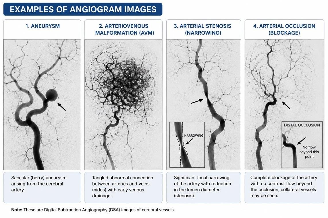
Cerebral angiograms are also called digital subtraction angiography of the brain (DSA).
Why is it Done?
- To diagnose brain aneurysms
- To detect stenosis (narrowing) or blockage of brain arteries
- To diagnose arteriovenous malformations (AVMs)
- To evaluate stroke or transient ischemic attack (TIA)
- To investigate unexplained brain bleeding
- To plan or guide brain treatments such as coiling, embolization, or stenting
- Vascular dissection (when the inside wall of an artery tears).
- To see how blood vessels are connected to or “feeding” a brain tumour.
- To learn more information about abnormalities which the doctor see on other imaging tests, such as an MRI or CT scan.
Benefits of Early Treatment for Cerebral Angiography:
Early diagnosis through cerebral angiography offers several advantages:
- Accurate Diagnosis: Provides detailed images of brain blood vessels to identify aneurysms, AVMs, and blockages.
- Timely Treatment: Early detection reduces the risk of complications like aneurysm rupture or stroke.
- Improved Outcomes: Early intervention increases the chances of successful treatment and recovery.
- Better Treatment Planning: Helps doctors devise the most effective treatment strategies.
Types of Angiography Used:
–The main types of angiography used to evaluate the brain are:
1. Digital Subtraction Angiography (DSA)/ Traditional Angiography
- Gold standard for imaging brain blood vessels.
- Performed by inserting a catheter into an artery (usually the femoral or radial artery).
- Contrast dye is injected directly into the blood vessels.
- Provides the most detailed images.
- Can be used for both diagnosis and treatment (e.g., coiling, embolization, stenting).
2. CT Angiography (CTA)
- Uses a CT scanner and contrast dye injected into a vein in the arm.
- No catheter is placed into the brain arteries.
- Fast and widely available.
- Commonly used in emergency situations, such as suspected stroke or aneurysm.
3. MR Angiography (MRA)
- Uses MRI to produce images of blood vessels.
- May be performed with or without contrast.
- No exposure to ionizing radiation.
- Useful for follow-up examinations and patients who should avoid radiation.
Pre-Procedure Evaluation:
- Medical history and physical examination
- Blood tests (kidney function-serum creatinine, complete blood count)
- Review of medications (especially blood thinners)
- Allergy history (especially contrast dye)
- Pregnancy test if needed
- CT scan or MRI of the brain, if indicated
Duration
About 30–60 minutes.
After the Procedure:
- You will be monitored for a few hours.
- Keep the puncture site straight as instructed.
- Drink plenty of fluids to help flush out the contrast dye.
- Avoid heavy lifting and strenuous activity for 24–48 hours.
- Follow your doctor’s instructions regarding medications.
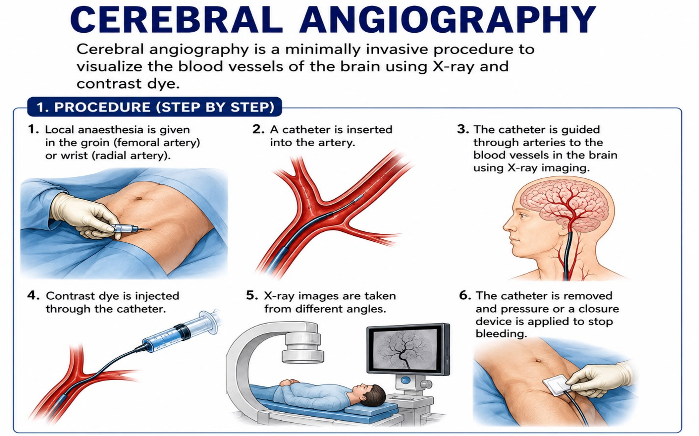
Procedure (Short Steps):
1. The patient lies in supine (flat on back) position. Local anaesthesia is given in the groin or wrist to take the puncture at the femoral or radial artery.
2. A catheter is inserted into the artery.
3. The catheter is guided to the arteries supplying the brain.
4. Contrast dye is injected.
5. X-ray images are taken from different angles.
6. The catheter is removed, and pressure or a closure device is used to stop bleed
Benefits:
- Provides highly detailed images of brain blood vessels.
- Helps diagnose vascular diseases accurately.
- Can guide treatment during the same procedure if needed.
Risks:
- Bleeding or bruising at the puncture site
- Allergic reaction to contrast dye
- Kidney injury from contrast dye if you have diabetes or kidney disease (rare)
- Stroke (rare)- If the catheter dislodges plaque from a vessel wall, it could block blood flow within your brain, causing a stroke
- Infection (very rare)
- Damage to the artery (rare)
When to Seek Medical Help?
Contact your doctor immediately if you develop:
- Severe headache
- Blurred vision
- Weakness, numbness, or difficulty speaking
- Chest pain, difficulty in breathing
- Heavy bleeding or swelling at the puncture site
- Severe pain, coldness, or discoloration of the arm or leg
- Fever or signs of infection such as warmth or excessive redness or discoloration.
FAQs
Is a cerebral angiogram considered surgery?
-A cerebral angiogram itself isn’t considered surgery. It’s a minimally invasive diagnostic procedure.
Would you like to request an appointment?
You can call on +91-98200 86520 for Appointments or fill the form below
If you’re experiencing symptoms of brain vascular issues, contact us today to schedule a consultation and explore your treatment options. Early diagnosis can prevent serious complications and improve your health outcomes.